Vaginal Septum: Symptoms, Types, and Treatment Options

Updated: 3 days ago
Medically reviewed by Dr. Baraa Alnahhal, MD · Last reviewed: October 17, 2026
Editorial note: This article is provided for educational purposes only. It is not medical advice. Always consult a qualified healthcare professional about your individual health situation.
Quick Answer
A vaginal septum is a congenital anomaly in which a band of tissue divides the vagina into two sections, forming when reproductive tubes fail to fuse properly during fetal development. [1] Symptoms often appear at puberty or early adulthood and include pelvic pain and painful sex. [1] Diagnosis uses a pelvic exam and imaging, and surgery can remove the tissue if needed. [1] [3]
What the research says
Outcomes after surgery are generally favorable but vary by anomaly type. A systematic review of 96 studies on surgical correction of obstructive genital tract anomalies found that, after repair of a transverse vaginal septum, 52.6% of women had infertility (pooled), compared with 8.0% after imperforate hymen repair. Across anomaly types, live-birth rates exceeded 80% among pregnancies achieved, preterm birth occurred in 23.7% of pregnancies and 55.9% of deliveries were cesarean. (Drever et al., Eur J Obstet Gynecol Reprod Biol, 2026) [2]
Timely diagnosis can prevent labor complications. A narrative review that searched four databases and identified 34 relevant articles (including 16 case reports on labor outcomes) concluded that diagnosis by pelvic examination and imaging such as MRI or ultrasound can prevent labor complications. Postoperative outcomes were generally favorable, but careful surgical planning is needed to avoid complications such as restenosis or infection. (Wu et al., Tzu Chi Med J, 2026) [3]
Ultrasound matched surgery findings in most obstructive Müllerian anomalies. In a pilot study of 29 women with surgically confirmed obstructive Müllerian anomalies, 3D-enhanced pelvic ultrasound agreed with the surgical diagnosis in 27 cases (93.1%), versus 24 cases (82.8%) for MRI. (Qin et al., J Pediatr Adolesc Gynecol, 2025) [4]
When to Call Your Care Team
Seek immediate medical attention if you experience any of the following, as they may indicate a complete blockage causing internal blood accumulation:
Sudden, severe pelvic pain
A complete inability to have a menstrual period despite other signs of puberty
Difficulty urinating
What Is a Vaginal Septum?
A vaginal septum occurs when a wall of tissue divides the vaginal canal, which is the muscular tube leading to the cervix and uterus. This anomaly happens during fetal development when the Müllerian ducts—two tubes that should fuse to form the vagina—fail to join properly. [1] Because the septum is internal, most people are unaware of the condition until they begin menstruating or become sexually active. [1]
Types of Vaginal Septums
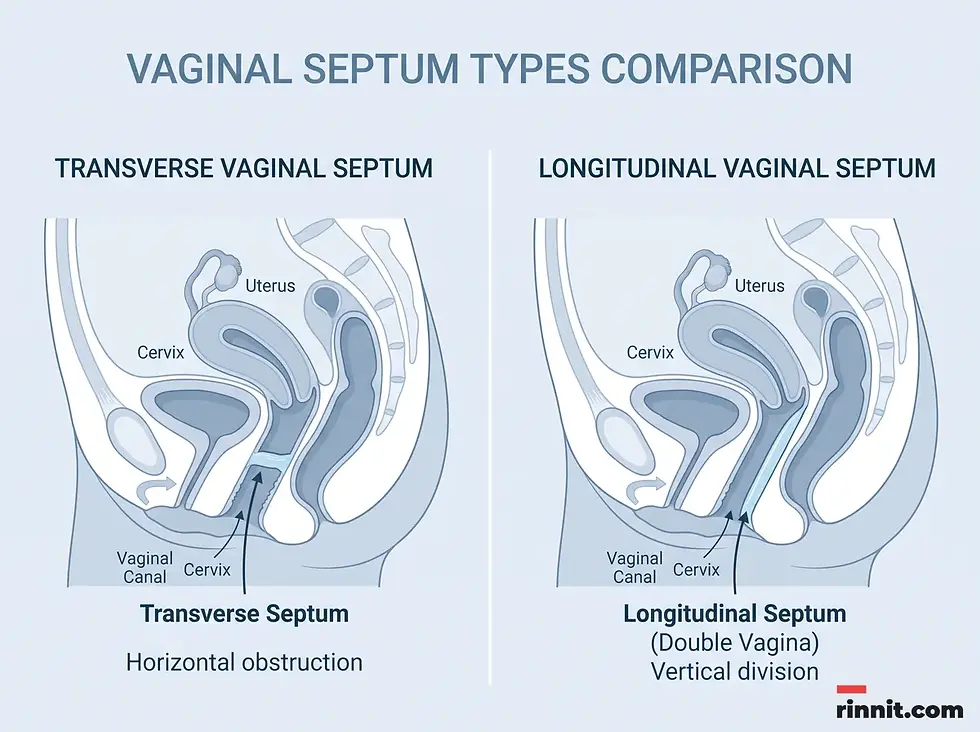
The classification of a vaginal septum depends on the direction of the tissue wall. Understanding the specific type is critical for determining the impact on menstruation and fertility.
Type | Direction | Description | Impact on Menstruation |
Transverse | Horizontal | Divides the vagina into top and bottom sections. [1] | Can block blood flow (complete) or allow slow flow (perforated). [1] |
Longitudinal | Vertical | Splits the vagina into two separate vertical cavities. [1] | Creates a “double vagina”; blood flows but tampons may leak. [1] |
Transverse Vaginal Septum
In this type, the tissue runs across the canal. It can be:
Complete: The tissue blocks the entire canal, preventing menstrual blood from exiting. This can cause blood to back up into the uterus, leading to significant pain and swelling. [1]
Perforated (partial): A small opening exists in the tissue, allowing blood to pass slowly. Periods may last longer than seven days due to the restricted flow. [1]
Longitudinal Vaginal Septum
Also called a vertical septum, this type creates two distinct vaginal canals. [1] It is frequently associated with other uterine anomalies, such as a didelphic uterus (two separate uteruses and cervixes). [1] While blood can exit normally, using tampons can be difficult because blood may leak from the cavity not containing the tampon. [1]
Common Symptoms
Many individuals do not realize they have a vaginal septum until a routine pelvic exam. [1] However, when symptoms occur, they usually relate to menstruation or sexual activity. [1]
Symptom | Description |
Amenorrhea | The complete absence of a menstrual period due to a total blockage. [1] |
Long periods | Menstruation lasting longer than seven days due to a perforated septum. [1] |
Pelvic pain | Abdominal discomfort caused by blood collecting in the upper vagina or uterus. [1] |
Tampon issues | Inability to insert a tampon, pain during use, or leaking despite proper placement. [1] |
Painful sex | Discomfort or pain during intercourse (dyspareunia). [1] |
Infertility | Difficulty conceiving due to anatomical obstructions. [1] |
Causes and Risk Factors
A vaginal septum is a congenital disorder, meaning it is present at birth. [1] Around six weeks into fetal development, the Müllerian ducts are supposed to fuse into a single canal. [1] A septum forms when this fusion process is incomplete. [1] Currently, there are no known risk factors or external causes for this developmental anomaly, and it is not considered preventable.
Diagnosis and Evaluation
Diagnosis typically begins during a physical pelvic exam. [1] [3] If a provider suspects a septum, they will use imaging tools to confirm the diagnosis and check for associated anomalies:
MRI (magnetic resonance imaging): Provides a detailed view of the tissue wall and reproductive tract. [1] [3]
Ultrasound: Helps identify the type of septum and any duplications in the uterus or cervix. [1]
Kidney/rectal screening: Providers may check these areas, as vaginal anomalies can sometimes be associated with issues in the kidneys or rectum. [1]

Treatment and Management
Treatment is generally not required if the septum is asymptomatic and does not affect fertility. [1] However, for most symptomatic cases, surgical intervention is the standard of care. [1]
Surgical Correction
Surgery involves the removal of the tissue wall to create a single, functional vaginal canal. [1]
Preparation: The procedure is performed in a hospital under general anesthesia. [1]
Tissue removal: The surgeon removes the obstructing wall and drains any trapped blood. [1]
Recovery: Once the tissue is removed, it does not grow back, although scar tissue can narrow the vagina again (restenosis) in some people, so follow-up visits matter. [1] The procedure improves menstrual flow, reduces pain during sex, and significantly lowers complications during future pregnancies and deliveries. [1]

Conclusion
A vaginal septum is a treatable anatomical condition that, while rare, can significantly impact a person’s quality of life and reproductive health. If you experience persistent pelvic pain, irregular menstruation, or discomfort during intercourse, consult a healthcare provider for a comprehensive evaluation. Modern surgical techniques offer a permanent solution, restoring normal anatomy and preventing long-term complications.
Frequently Asked Questions
1. Is a vaginal septum a common condition?
No, it is very rare, occurring in less than 0.03% of infants.
2. Can I get pregnant if I have a vaginal septum?
Yes, conception is possible, but the septum can increase the risk of complications. Fixation is recommended before pregnancy.
3. Does a vaginal septum cause cancer?
No, a vaginal septum is a benign (non-cancerous) band of tissue.
4. Will I need surgery if I don’t have symptoms?
Not necessarily. If it doesn’t cause pain or affect your health, your provider may suggest simple monitoring.
5. Can a vaginal septum be seen from the outside?
No, the tissue wall is internal and can only be identified during a pelvic exam or via imaging.
6. What is a “double vagina”?
A “double vagina” is another term for a longitudinal vaginal septum, where the vagina is split into two separate canals.
7. Can a vaginal septum tear on its own?
Yes, in some cases, the tissue may tear naturally during sexual activity, which may eliminate the need for surgery.
8. Is the surgery to remove a septum painful?
The surgery is performed under general anesthesia, so you won’t feel pain during the procedure. Post-operative discomfort is managed with medication.
9. Can a vaginal septum grow back after surgery?
The septum itself does not regrow once removed, but scar tissue can sometimes narrow the vagina again (restenosis), so follow-up with your surgeon is important.
10. How long does the surgery take?
The procedure typically takes a few hours in a hospital setting.
11. Are there different types of transverse septums?
Yes, they can be complete (blocking all flow) or perforated (allowing some flow through a small hole).
12. Why do tampons leak if I have a longitudinal septum?
Because there are two canals, a tampon in one canal will not stop blood flowing from the other cavity.
13. Does a vaginal septum affect my kidneys?
Sometimes. Vaginal anomalies can be associated with kidney malformations, so your doctor may check your renal health.
14. When do symptoms usually start?
Most people notice symptoms during puberty (when periods start) or when they become sexually active.
15. Can an ultrasound detect a vaginal septum?
Yes, ultrasounds are commonly used to confirm the diagnosis and identify the type of septum.
Related Reading
References
Cleveland Clinic. Vaginal septum. Cleveland Clinic. January 13, 2023. https://my.clevelandclinic.org/health/diseases/24677-vaginal-septum
Drever N, Gane C, Bailey M. Reproductive outcomes after surgical correction of congenital obstructive genital tract anomalies: a systematic review and quantitative pooled analysis of published data. Eur J Obstet Gynecol Reprod Biol. 2026;320:115014. PMID 41713152. https://doi.org/10.1016/j.ejogrb.2026.115014
Wu YC, Li PC, Ding DC. Vaginal septum in women: a review of diagnosis, management, and obstetric outcomes. Tzu Chi Med J. 2026;38(1):40-47. PMID 41669329. https://doi.org/10.4103/tcmj.TCMJ-D-25-00041
Qin C, Lee P, Luo L. Comparison between 3D-enhanced conventional pelvic ultrasound and magnetic resonance imaging in the evaluation of obstructive Müllerian anomalies and its concordance with surgical diagnosis. J Pediatr Adolesc Gynecol. 2025;38(2):174-179. PMID 39098548. https://doi.org/10.1016/j.jpag.2024.07.004



Comments