Understanding the Ankle Joint: Anatomy, Function, and Common Conditions

Updated: 2 days ago
Medically reviewed by Dr. Baraa Alnahhal, MD · Last reviewed: October 2026
Quick Answer: What is the ankle joint?
The ankle joint, medically called the tibiotalar or talocrural joint, is the synovial hinge joint connecting the foot and lower leg. Formed by the tibia, fibula and talus bones and supported by cartilage, ligaments and muscles, it allows plantar flexion and dorsiflexion. Common conditions include arthritis, sprains and fractures, and the RICE method helps manage pain and swelling.
What the research says
According to PubMed, two recent systematic reviews address how ankle sprains, the most common ankle injury, are best managed:
Clinical practice guidelines: A PRISMA systematic review of seven guidelines found that the Ottawa rules, manual therapy, cryotherapy, functional supports, early ambulation, short-term NSAIDs, and rehabilitation had enough evidence to be highly recommended for ankle sprain management. DOI · PMID 36281183
Umbrella review: An umbrella review of 24 systematic reviews found evidence supporting non-surgical treatment for acute ankle sprain, with functional treatment preferable to immobilization, and supervised exercise therapy helping to prevent re-injury and restore ankle dorsiflexion. DOI · PMID 35872766
What is the ankle joint?
The ankle joint is the primary connection between the foot and the lower leg [1]. In medical contexts, healthcare providers may refer to it as the tibiotalar joint or the talocrural joint [1]. As a vital component of the skeletal system, the ankle enables essential movements required for walking, running, and maintaining balance [1].
What does the ankle joint do and how does it move?
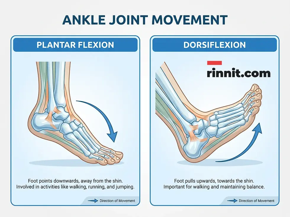
The primary role of the ankle joint is to provide stability and flexibility during physical activity [1]. It moves in two specific directions to maintain balance and facilitate locomotion:
Plantar Flexion: Moving the foot downward, away from the body [1].
Dorsiflexion: Moving the foot upward, toward the body [1].
What is the anatomy and structure of the ankle joint?
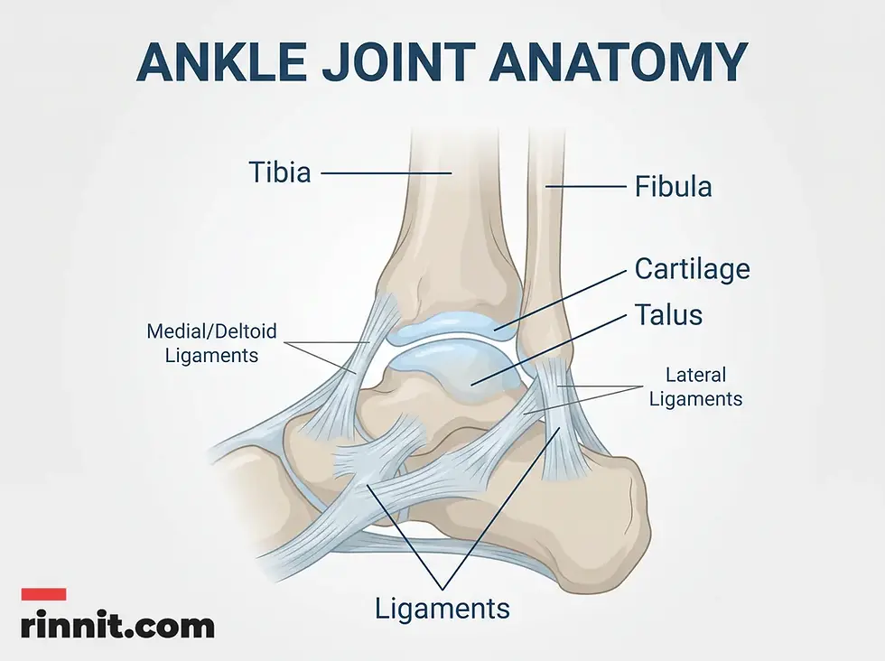
The ankle is classified as a synovial joint, which offers the greatest range of motion among joint types [1]. It features a cavity lined with a synovial membrane that secretes lubricating fluid to reduce friction during movement [1]. Functionally, it acts as a hinge joint, similar to a door hinge, moving a specific distance to open and close [1].
Skeletal Composition
Three primary bones meet to form the ankle joint:
Tibia: The larger shin bone on the inner side of the leg [1].
Fibula: The thinner calf bone on the outer side of the leg [1].
Talus: The bone located between the heel bone and the tibia/fibula [1].
Connective Tissues
The joint is protected and stabilized by several layers of tissue:
Cartilage: Strong, smooth hyaline cartilage (articular cartilage) caps the ends of the bones, acting as a shock absorber and allowing bones to glide smoothly [1].
Ligaments: Cords of tissue that connect the bones. These include the medial (deltoid) ligaments on the inside, lateral ligaments on the outside, and syndesmotic ligaments connecting the tibia and fibula [1].
Muscles, Nerves, and Blood Supply
Multiple muscles in the leg and foot control ankle movement, including the gastrocnemius and soleus for downward flexion, and the tibialis anterior for upward movement [1]. Nerves like the tibial and peroneal nerves carry impulses for sensation and motion, while the tibial and peroneal arteries provide the necessary blood supply [1].
Component Category | Key Structures in the Ankle |
Bones | Tibia, Fibula, Talus [1] |
Ligaments | Medial (Deltoid), Lateral, Syndesmotic [1] |
Nerves | Tibial, Superficial Peroneal, Deep Peroneal [1] |
Arteries | Anterior Tibial, Posterior Tibial, Peroneal [1] |
What are common ankle joint conditions and disorders?
Because the ankle is used constantly, it is prone to both acute injuries and chronic conditions [1]. Common issues include:
Arthritis: Inflammation of the joint, leading to pain and stiffness [1].
Gout: A type of arthritis caused by the buildup of uric acid crystals [1].
Bursitis and Tendinitis: Inflammation of the fluid-filled sacs (bursae) or tendons [1].
Sports Injuries: Including ankle sprains, fractures, and Achilles tendon ruptures [1].
What are the symptoms of ankle joint problems and how are they diagnosed?
Symptoms of an ankle issue typically depend on the underlying cause but often include pain, swelling (inflammation), stiffness, and instability [1]. In some cases, redness or a feeling of warmth may be present [1].
To diagnose these conditions, healthcare providers utilize various tools:
Imaging: X-rays, ultrasounds, CT scans, and MRIs to view bones and soft tissues [1].
Lab Tests: Blood tests or joint aspiration (removing fluid for testing) to identify infection or gout [1].
How do you care for and manage ankle joint pain?
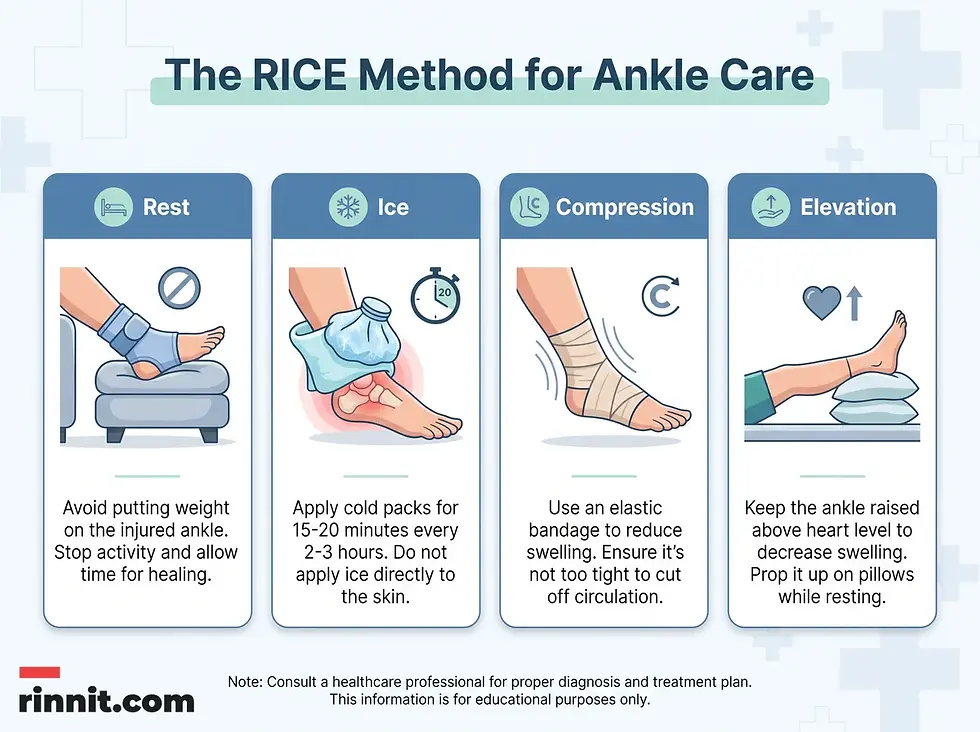
Minor ankle pain or swelling can be relieved right away with the RICE method: rest, ice, compression and elevation.
Rest: Avoid activities that cause pain or stress the joint [1].
Ice: Apply cold packs for 15 minutes several times a day [1].
Compression: Use an elastic bandage to minimize swelling [1].
Elevation: Keep the ankle propped above heart level [1].
Conclusion
The ankle joint is a complex and essential structure that facilitates mobility and balance. By understanding its anatomy—from the bones and ligaments to the muscles that drive movement—individuals can better recognize the signs of injury or chronic conditions. Early intervention and proper at-home care are key to preserving joint health and maintaining an active lifestyle.
If you are experiencing severe ankle pain, persistent swelling, or instability that does not improve with rest, consult a healthcare professional. A proper diagnosis and personalized treatment plan can help prevent long-term complications and ensure you return to your daily activities safely.
Frequently Asked Questions
What is the ankle joint?
The ankle joint, also known as the tibiotalar or talocrural joint, is the connection between the foot and the lower leg [1].
Which bones make up the ankle?
The ankle is formed by three bones: the tibia (shin bone), the fibula (calf bone), and the talus [1].
What type of joint is the ankle?
The ankle is a synovial hinge joint, which allows for smooth movement in two main directions [1].
What are the primary movements of the ankle?
The ankle moves in two directions: plantar flexion (downward) and dorsiflexion (upward) [1].
What is the role of cartilage in the ankle?
Hyaline cartilage caps the ends of the bones, acting as a shock absorber and ensuring smooth movement with minimal friction [1].
What are the different types of ankle ligaments?
The ankle has three main sets of ligaments: medial (deltoid) ligaments, lateral ligaments, and syndesmotic ligaments [1].
What are common symptoms of an ankle injury?
Common symptoms include pain during movement, swelling, stiffness, instability, and redness or warmth around the joint [1].
How is an ankle injury diagnosed?
Diagnosis typically involves physical examination and imaging tests such as X-rays, MRIs, CT scans, or ultrasounds [1].
What is the RICE method for ankle care?
The RICE method stands for Rest, Ice, Compression, and Elevation, which helps manage pain and swelling after an injury [1].
When should I see a doctor for ankle pain?
You should consult a healthcare provider if pain is severe, if there is significant instability, or if symptoms do not improve after a few days of rest [1].
Related Reading
Ankle Arthroscopy: Procedure, Risks and Recovery explains ankle arthroscopy and recovery.
Ankle Replacement Surgery: Procedure, Risks and Recovery explains ankle replacement surgery for arthritis.
References
[1] Cleveland Clinic. Ankle Joint. Cleveland Clinic Health Library. Last updated April 15, 2023. https://my.clevelandclinic.org/health/body/24909-ankle-joint
[2] Ruiz-Sánchez FJ, Ruiz-Muñoz M, Martín-Martín J, et al. Management and treatment of ankle sprain according to clinical practice guidelines: a PRISMA systematic review. Medicine (Baltimore). 2022;101(42):e31087. PMID 36281183. https://doi.org/10.1097/MD.0000000000031087
[3] Gaddi D, Mosca A, Piatti M, et al. Acute ankle sprain management: an umbrella review of systematic reviews. Front Med (Lausanne). 2022;9:868474. PMID 35872766. https://doi.org/10.3389/fmed.2022.868474
Medical disclaimer
The information provided in this article is for educational purposes only and should not be used as a substitute for professional medical advice, diagnosis, or treatment. Always seek the advice of your physician or other qualified health provider with any questions you may have regarding a medical condition.



Comments