Understanding Nipple Anatomy, Function, and Health

Updated: 3 days ago
Medically reviewed by Dr. Baraa Alnahhal, MD · Last reviewed: October 17, 2026
The nipple is a small structure at the center of the breast that is essential for breastfeeding and also provides sensation. Together with the surrounding areola it forms the nipple-areolar complex. Flat or inverted nipples are often harmless traits you are born with, but sudden changes in appearance, texture, or sensation can point to conditions such as eczema or breast cancer, which is why regular screening and prompt evaluation of changes matter [1].
Quick Answer: What is the nipple?
The nipple is the small structure at the center of the breast, surrounded by a darker area called the areola. It contains smooth muscle, nerves and milk duct openings, and it delivers milk during breastfeeding and provides sensation. Nipples may protrude, be flat or inverted, but discharge, a rash or a new inversion needs medical evaluation to rule out cancer. [1]
What the research says
Screening guidelines differ on how often to get a mammogram. The US Preventive Services Task Force recommends mammography every two years for women aged 40 to 74 (moderate certainty of moderate net benefit) and found the evidence insufficient for women 75 and older and for supplemental ultrasound or MRI in women with dense breasts, while Cleveland Clinic advises annual mammograms starting at 40 (USPSTF, JAMA, 2024 [2]).
A mammogram alone can miss cancer in people with nipple discharge. A network meta-analysis of 36 studies including 3,764 patients with pathologic nipple discharge found sensitivity for detecting cancer was 83% for MRI, 58% for ductoscopy, 50% for ultrasound, 38% for cytology, and only 22% for mammography (specificity 93%), which is why discharge needs a full evaluation (Filipe et al., Clin Breast Cancer, 2020 [3]).
What is the anatomy of the nipple and how does it vary?
The nipple and the surrounding darker skin, the areola, together form the nipple-areolar complex (NAC). On average, a nipple is 10 to 12 millimeters wide and sticks out 9 to 10 millimeters from the surrounding skin. It is made up of smooth muscle fibers, connective tissue, and milk ducts, which are tubes that carry milk. Each nipple has about nine small openings where milk comes out, and hundreds of nerves provide sensation [1].
Nipple appearance varies significantly. Most people have two nipples, but some have extra nipples (supernumerary nipples), and some are born without any (athelia), which is sometimes associated with a genetic disorder such as Poland syndrome [1].
Nipple type | Description | Frequency and context |
Protruding | Sticks outward from the areola, like a tiny cylinder | The most common type |
Flat | Level with the skin of the areola | Often a harmless variation you are born with |
Inverted | Points inward, tucked inside the areola | Often present from birth; a sudden change needs review |
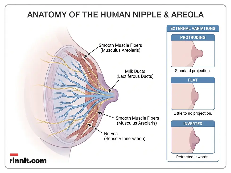
The nipple contains smooth muscle fibers, milk ducts, and nerves, and can be protruding, flat, or inverted.
Flat or inverted nipples are often harmless variations. But if you have protruding nipples and suddenly notice they are flat or inverted, tell a healthcare provider: the change can be harmless, but it is sometimes a sign of cancer or another condition that needs treatment [1].
What is the function of the nipple?
The most vital role of the nipple is in breastfeeding. During nursing, a baby latches onto the nipple and areola, and tiny openings in the nipple release milk to nourish the baby. Nipples are also a source of sexual pleasure for all people because of their many nerves [1].
The smooth muscle fibers in the nipple contract in response to stimuli such as cold or touch, which makes the nipple more prominent.
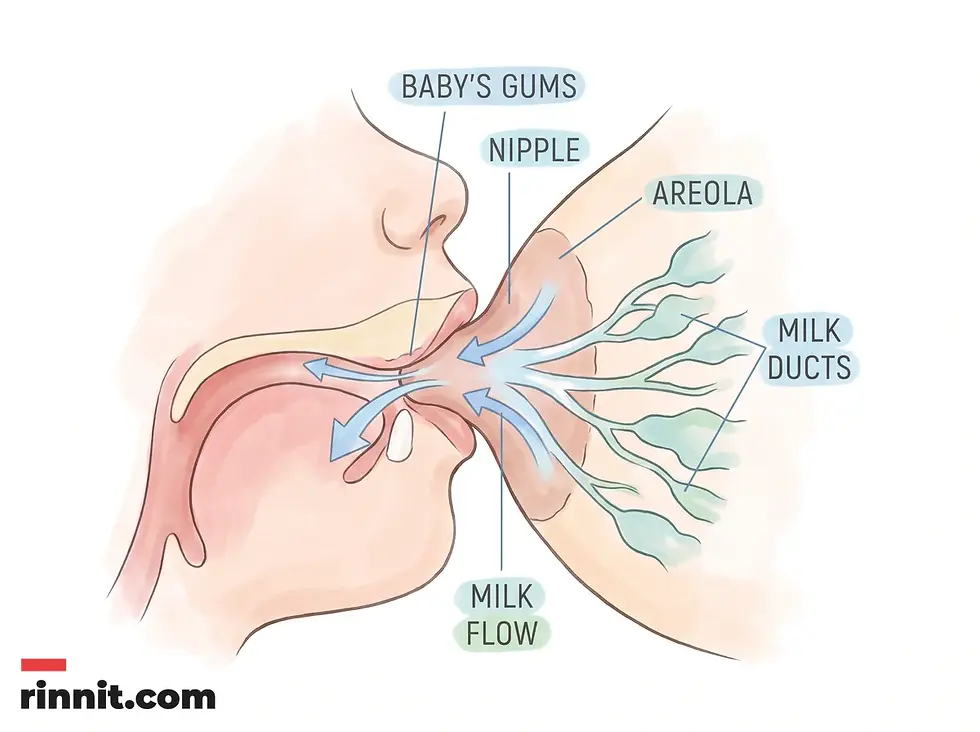
During breastfeeding, the baby latches onto the nipple and areola and milk flows from the milk ducts through the nipple.
What conditions affect the nipple and how are they screened?
Nipple conditions range from harmless rashes like nipple eczema to cancers such as Paget's disease of the breast, ductal carcinoma in situ and invasive ductal carcinoma. Breast cancer can affect the whole breast, including the nipple. Types that start in the nipple area include [1]:
Paget's disease of the breast: Cancer that develops in the skin of the nipple, causing itching, burning, discharge, or a rash on the nipple or areola.
Ductal carcinoma in situ (DCIS): Cancer that starts in the milk ducts and typically does not spread beyond them. It usually causes no symptoms, and most cases are found on imaging.
Invasive ductal carcinoma (IDC): Cancer that starts in the milk ducts and spreads to nearby tissue. Symptoms may include dimpled, puckered, scaly, or inflamed skin on the breast or nipple, a lump, or changes in breast size or shape.
Nipple eczema, an itchy, red, scaly rash on the nipple and areola, can also affect the nipples. It is not always easy to tell eczema from signs of breast cancer, which is why it is vital to call a provider if you notice any change [1].
Healthcare providers routinely use mammograms to check the whole breast, including the nipple, and use breast ultrasound and breast MRI in certain situations [1].
Screening tool | Primary use | Recommended frequency |
Mammogram | X-ray imaging of breast tissue | Cleveland Clinic: annually from age 40; USPSTF: every two years from 40 to 74; sooner if your risk is higher |
Breast ultrasound | Uses sound waves to check specific lumps | As needed for follow-up |
Breast MRI | Detailed imaging in certain situations, such as high risk | Supplemental to mammograms |
Males can also develop breast cancer, which is rare; if you are male, your provider may recommend screening if you have certain risk factors [1].

Nipple and breast care combines awareness of what is normal for you, regular screening, and prompt evaluation of warning signs.
Conclusion
Though small, the nipple is a complex structure with important biological and sensory roles. Most variations in its appearance are natural and harmless, but staying alert to change is key to long-term health. Combining breast awareness with professional screening helps protect against serious conditions and keeps the nipple-areolar complex healthy [1].
What steps can you take for nipple health?
Nipple health starts with monthly self-exams, following breast screening guidelines, contacting a provider about red flags such as discharge or new inversion, and caring properly for piercings.
Monthly self-exams: Get to know the normal look and feel of your breasts and nipples so you can notice changes early [1].
Follow screening guidelines: Cleveland Clinic advises annual mammograms starting at age 40, while the USPSTF recommends every two years from 40 to 74; ask your provider about the schedule that fits your risk [1] [2].
Watch for red flags: Contact a healthcare provider if you notice nipple pain that lasts several days, changes in the color or texture of the skin, cracks, a rash, discharge, or a nipple that has newly become flat or inverted [1].
Protect the skin: If you have a nipple piercing, ask your provider how to care for it, including any precautions during pregnancy or breastfeeding [1].
Frequently Asked Questions
What is the nipple-areolar complex?
The nipple-areolar complex refers to the nipple and the surrounding darker-colored skin called the areola.
What is the primary function of the nipple?
The nipple's main role is to release milk during breastfeeding to nourish a baby.
Are flat or inverted nipples normal?
Yes, many people are born with flat or inverted nipples, and they are typically harmless variations.
What should I do if my protruding nipple suddenly becomes inverted?
You should contact a healthcare provider, as sudden changes can sometimes indicate an underlying condition like cancer.
How many milk ducts are in a nipple?
Each nipple typically has about nine small openings where milk is released.
Can men get breast cancer?
Yes. While rare, breast cancer can affect males and may cause changes to the nipple area.
What is Paget's disease of the breast?
Paget's disease of the breast is a type of cancer that develops in the skin of the nipple, often causing itching, burning, discharge, or a rash.
When should I start getting mammograms?
Cleveland Clinic recommends starting annual mammograms at age 40, and the US Preventive Services Task Force recommends mammograms every two years from ages 40 to 74. Your provider may recommend starting sooner depending on your risk, so ask which schedule fits you.
What causes nipple discharge?
Discharge can be caused by various factors, ranging from normal lactation to infections or cancer, and it should always be evaluated by a healthcare professional.
How do I perform a breast self-exam?
Check your breasts and nipples once a month so you know how they normally look and feel, and tell a healthcare provider about any new lumps, skin changes, rash, discharge, or persistent soreness.
Related Reading
Flat and Inverted Nipples: Causes, Breastfeeding, and Care explains flat and inverted nipples and tips for breastfeeding.
Cracked Nipples (Nipple Fissures): Symptoms, Causes, Treatment and Healing — What You Need to Know covers why nipples crack and how to help them heal.
Mastitis: Symptoms, Causes, and Treatment describes breast tissue inflammation and infection and how it is treated.
Paget's Disease of the Breast: Symptoms, Causes, and Treatment — A Clear Guide to Understanding and Managing the Condition explains a rare breast cancer that starts on the nipple.
References
Cleveland Clinic. Nipple: Anatomy & Types. Cleveland Clinic. January 7, 2025. https://my.clevelandclinic.org/health/body/nipple
US Preventive Services Task Force; Nicholson WK, Silverstein M, Wong JB, et al. Screening for breast cancer: US Preventive Services Task Force recommendation statement. JAMA. 2024;331(22):1918-1930. PMID: 38687503. DOI: 10.1001/jama.2024.5534
Filipe MD, Patuleia SIS, de Jong VMT, et al. Network meta-analysis for the diagnostic approach to pathologic nipple discharge. Clin Breast Cancer. 2020;20(6):e723-e748. PMID: 32665191. DOI: 10.1016/j.clbc.2020.05.015
Medical disclaimer
This article is for informational purposes only and is not a substitute for professional medical advice, diagnosis, or treatment. The clinical information in this guide reflects source material last reviewed in January 2025.



Comments