Sural Nerve: Anatomy, Essential Functions, and Care

Updated: 3 days ago
Medically reviewed by Dr. Baraa Alnahhal, MD · Last reviewed: October 2026
The sural nerve is a sensory nerve just below the skin of the back of the lower leg. Formed where the tibial and common fibular nerves join, it carries touch, temperature, pain, vibration and foot-position signals from the back of the leg below the knee, the outer foot, outer heel and ankle [1]. It is also a source of tissue for nerve biopsies and grafts, and diabetes, sports injuries, entrapment and surgery can affect it [1].
Quick Answer
The sural nerve is a sensory nerve just below the skin in the back of the lower leg that lets you detect touch, foot position, temperature, pain and vibration. It is part of the peripheral nervous system. Because of its length and ability to regenerate, providers also use it for nerve biopsies and grafts to manage complex nerve problems.
What the research says
Taking a sural nerve graft usually leaves some numbness. A systematic review of 9 studies (240 patients) found that 87.2% reported sensory loss after sural nerve harvest, 25.6% pain, 22.2% cold sensitivity and 10% functional impairment; sparing the proximal sural nerve was linked to less pain (9.1% vs 35.5%) than harvesting at the back of the knee (Bamba et al., Journal of Plastic, Reconstructive & Aesthetic Surgery, 2021 [2]).
The sural nerve is not the best donor for every graft. A systematic review of digital (finger) nerve grafting found acceptable donor-site morbidity for the sural nerve (15%) but the lowest sensory recovery of the donors compared (46% good to very good recovery), and proposed matching the donor nerve to the injury zone (Kang, Annals of Plastic Surgery, 2025 [3]).
Blood sugar control tracks with sural nerve health in diabetes. In 424 people with diabetic neuropathy followed for 52 weeks, baseline HbA1c was the only factor that differed between those whose sural nerve fibers regenerated (8.3%) and those whose fibers degenerated (9.2%) (odds ratio 0.68, 95% CI 0.54 to 0.85), and the authors conclude that optimal blood glucose control is likely essential to prevent continued nerve injury (Hur et al., Diabetes Care, 2013 [4]).
What is the anatomy and function of the sural nerve?
Your sural nerve starts in the upper calf, where the tibial nerve and the common fibular (common peroneal) nerve join. It travels down the back outer part of your leg, curves at your ankle and ends before your toes [1]. The tibial nerve enables sensation and movement in the back of the leg and foot, and the common fibular nerve provides sensory and movement capabilities to the area behind the knee and nearby leg tissue.
The sural nerve contains tiny string-like fibers (axons) bound into bundles by a fatty substance called myelin. These bundles connect up to your spinal cord, which lets nerve signals travel quickly to and from your brain [1].
What it detects | Why it matters |
Touch | Skin sensation on the back of the leg below the knee, the outer foot, the outer heel and the ankle |
Temperature and pain | Warns you about hot, cold and harmful sensations in the same areas |
Vibration | Lets you sense vibration against the skin of the lower leg and foot |
Foot position | Contributes to maintaining balance while you are on your feet |

What are sural nerve biopsies and grafts?
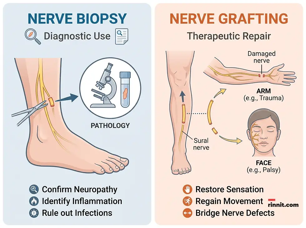
A sural nerve biopsy takes a tissue sample that healthcare providers examine in a lab for signs of nerve disease. The sural nerve is used because it lies close to the skin's surface and is easy to find, and serious complications are uncommon, though it usually leaves a lasting patch of numbness on the outer foot [1]. You may need a biopsy if you show signs of AL (light chain) amyloidosis, vasculitis (inflammation in blood vessels) or any peripheral nerve disorder that is difficult to diagnose [1].
A nerve graft uses healthy tissue to repair or replace damaged nerves, and the sural nerve is chosen because it provides a long piece of tissue [1]. A sural nerve graft can be attached elsewhere in the body to relieve:
Brachial plexus injuries, which affect the shoulder, arm or hand.
Sciatic or peroneal nerve injuries, which affect the leg, ankle or foot.
Erectile dysfunction.
Facial paralysis, nerve damage that may cause one side of the face to sag.
Neurotrophic keratitis, a rare disease affecting nerves in the eye.
What common conditions affect the sural nerve?
Conditions that affect the sural nerve include diabetes-related neuropathy, nerve entrapment, sports injuries such as ankle sprains and surgical complications.
Diabetes-related neuropathy: Nerve damage from ongoing high blood sugar. The sural nerve is one of the most commonly affected nerves [1].
Sural nerve entrapment (pinched nerve): Nearby tissue thickens and presses on the nerve, causing sural nerve pain [1].
Sports injuries: Ankle sprains can damage the nerve, and so can ski boots or horse saddle ankle straps that are too tight [1].
Surgical complications: Surgical instruments can injure the nerve, and hardware such as screws used to repair a bad break can rub against it [1].
When should you call a healthcare provider about sural nerve symptoms?
Call a healthcare provider about your sural nerve if you have discomfort, burning, numbness or tingling in your lower leg or the top outside of your foot. Symptoms may include:
Burning.
Lack of sensation.
Sensitivity to touch.
Sharp or throbbing pain.
Tingling or numbness.
How can you protect your sural nerve?
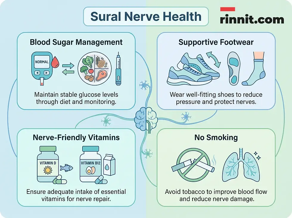
Protecting your sural nerve means controlling blood sugar if you have diabetes, wearing well-fitting athletic shoes, quitting tobacco and eating foods with vitamins D and B12.
Keep blood sugar in a healthy range if you have diabetes.
Choose well-fitting athletic footwear and replace it once it wears out.
Quit smoking or using other tobacco products.
Eat a healthy diet with foods that contain vitamin D and vitamin B12, which support nerve health.
Conclusion
The sural nerve gives you sensation in the calf, ankle and outer foot, helps your balance, and is valuable to doctors for diagnosing and repairing nerve problems. Protecting it starts with managing blood sugar, wearing well-fitting shoes, not smoking and eating a healthy diet.
Next step: If you have burning, numbness, tingling or sharp pain in your lower leg or outer foot, see your healthcare provider.
Frequently Asked Questions
What is the primary function of the sural nerve?
The sural nerve provides skin sensation to the back of your leg below the knee, the outer side of your foot, your outer heel and your ankle.
Where exactly is the sural nerve located?
The sural nerve lies just below the skin in the back of your lower leg (calf). It travels down the back outer part of the leg, curves at the ankle and ends before the toes.
What nerves form the sural nerve?
The sural nerve starts in the upper calf where two nerves join: the tibial nerve and the common fibular (common peroneal) nerve.
Can the sural nerve help with balance?
Yes. It lets you detect foot position and contributes to maintaining balance while you are on your feet.
Why is the sural nerve used for biopsies?
The sural nerve lies close to the skin's surface and is easy to find, and serious complications are uncommon, though it usually leaves a lasting patch of numbness on the outer foot. A biopsy may be needed for signs of AL (light chain) amyloidosis, vasculitis or any peripheral nerve disorder that is difficult to diagnose.
What is sural nerve grafting?
A nerve graft uses healthy tissue to repair or replace damaged nerves. The sural nerve is used because it provides a long piece of tissue, and it can be attached elsewhere in the body to treat conditions such as brachial plexus injuries, sciatic or peroneal nerve injuries, facial paralysis and neurotrophic keratitis. Taking the nerve commonly leaves some numbness on the outer foot.
What are the symptoms of sural nerve damage?
Symptoms in the lower leg or the top outside of the foot can include burning, lack of sensation, sensitivity to touch, sharp or throbbing pain, and tingling or numbness. Call your healthcare provider if you have them.
How does diabetes affect the sural nerve?
Ongoing high blood sugar can cause diabetes-related neuropathy, and the sural nerve is one of the most commonly affected nerves. Keeping blood sugars within a healthy range is an important part of protecting it.
What is sural nerve entrapment?
Entrapment, or a pinched nerve, happens when nearby tissue thickens and presses on the nerve, causing sural nerve pain.
Which vitamins support sural nerve health?
Cleveland Clinic recommends a healthy diet with foods that contain vitamin D and vitamin B12, which support nerve health.
Related Reading
Peripheral Neuropathy: Symptoms, Causes, and Treatment Options Explained explains nerve damage that often first affects the feet and legs.
Nerve Conduction Study: What It Is, Procedure and Results describes the test that measures how well nerves carry signals.
Diabetic Neuropathy: Symptoms, Types, Causes & Treatment covers how diabetes damages nerves and how to protect them.
EMG (Electromyography): What It Tests, What to Expect and Results explains a test of muscle and nerve electrical activity.
References
Cleveland Clinic. Sural Nerve. Cleveland Clinic. Last updated January 18, 2022. https://my.clevelandclinic.org/health/body/22323-sural-nerve
Bamba R, Loewenstein SN, Adkinson JM. Donor site morbidity after sural nerve grafting: a systematic review. Journal of Plastic, Reconstructive & Aesthetic Surgery. 2021;74(11):3055-3060. PMID 33985927. https://doi.org/10.1016/j.bjps.2021.03.096
Kang D. Beyond one-size-fits-all: pioneering a zone-based strategy in digital nerve autografting. Annals of Plastic Surgery. 2025;94(6):663-669. PMID 39874557. https://doi.org/10.1097/SAP.0000000000004242
Hur J, Sullivan KA, Callaghan BC, et al. Identification of factors associated with sural nerve regeneration and degeneration in diabetic neuropathy. Diabetes Care. 2013;36(12):4043-4049. PMID 24101696. https://doi.org/10.2337/dc12-2530
Medical disclaimer
The information provided in this article is for educational purposes only and should not be used as a substitute for professional medical advice, diagnosis, or treatment. Always seek the advice of your physician or other qualified health provider with any questions you may have regarding a medical condition.



Comments