Skin Cancer on the Ear: Detection, Types, and Treatment

Updated: 3 days ago
Medically reviewed by Dr. Baraa Alnahhal, MD · Last reviewed: October 2026
Quick Answer: What is skin cancer on the ear?
Skin cancer on the ear is an uncontrolled growth of abnormal skin cells, typically caused by UV damage from the sun or tanning beds, with squamous cell carcinoma the most frequent type on the outer ear. [1] Signs include non-healing sores, changing moles or scaly pink lumps. [1] Biopsy confirms it, and treatment often involves surgery such as Mohs surgery. [1]
What the research says
Daily sunscreen lowered the number of squamous cell skin cancers. In a randomized trial of 1,621 adults in Queensland, Australia, daily SPF 15-plus sunscreen on the head, neck, arms, and hands led to significantly fewer squamous cell carcinoma tumours over 4.5 years (1,115 vs 1,832 per 100,000; rate ratio 0.61, 95% CI 0.46 to 0.81). It had no effect on basal cell carcinoma (Green et al., Lancet, 1999) [2].
Mohs surgery gives good local control of ear squamous cell carcinoma. In 170 invasive squamous cell carcinomas of the ear treated with Mohs surgery (mean follow-up 48 months), the local recurrence rate was 4.1% and the rate of lymph node metastasis was 10.6%. Five-year disease-specific survival was 89.7%. Most patients were older men (93.2%), and the study was not randomized (González et al., J Plast Reconstr Aesthet Surg, 2021) [3].
The ear is a recognized high-risk site for squamous cell carcinoma. A review notes that most cutaneous squamous cell carcinomas are treated successfully with surgery, but local and distant metastases develop in about 5% of cases. The risk is higher with certain features, including a location on the pinna of the ear (Nuño-González et al., Actas Dermosifiliogr, 2012) [4].
What is skin cancer on the ear?
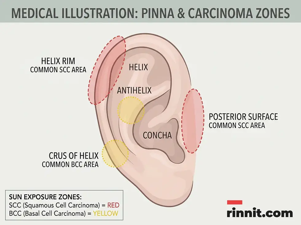
Skin cancer on the ear refers to the uncontrollable growth of abnormal cells, usually beginning on the outer portion of the ear known as the pinna. This area is particularly vulnerable because it is often neglected during sunscreen application despite being one of the most sun-exposed parts of the body. [1] If left undiagnosed, these cancerous cells can invade deeper tissues or travel through the blood and lymph systems to distant organs, including the brain. [1]
There are three primary types of skin cancer that affect the ear. Squamous cell carcinoma is the most common variety found in this location. [1] Basal cell carcinoma also frequently occurs and is generally slow-growing. Melanoma, while less common, is the most dangerous form due to its high potential for metastasis (see our guide to amelanotic melanoma). [1] Understanding the specific type of cancer is crucial for determining the most effective treatment strategy and long-term outlook. For other growths of the ear, see our overview of benign and cancerous ear tumors.
What are the symptoms of skin cancer on the ear?
The most frequent sign of ear skin cancer is a visible change in the skin’s appearance. [1] This might manifest as a sore that bleeds, itches, or fails to heal within four weeks. [1] Other common indicators include discolored patches of skin, shiny bumps, or yellow, scar-like areas. [1] Because the ear has many folds and is difficult to see directly, using a mirror or camera to perform regular checks is highly recommended.
For moles specifically, the ABCDE guideline serves as a vital tool for identification. Asymmetry in shape, irregular or blurred borders, and the presence of multiple colors within a single mole are all red flags. Additionally, any mole with a diameter larger than six millimeters or one that is evolving in size, shape, or color should be evaluated by a medical professional immediately. [1]
Sign Category | Specific Indicators | Visual Characteristics |
Growth type | Nodules or lumps | Shiny bumps or pink, scaly surfaces |
Surface changes | Non-healing sores | Bleeding, crusting, or itching spots |
Discoloration | Pigmentation changes | Yellow, white, or multi-colored areas |
Mole evolution | ABCDE changes | Increasing size or irregular borders |
What causes skin cancer on the ear, and who is at risk?
The overwhelming majority of ear skin cancer cases are caused by overexposure to ultraviolet (UV) light. [1] UV rays from the sun or tanning beds damage the DNA within skin cells, leading to mutations that cause cells to divide rapidly and form a malignant mass. [1] In some instances, long-term contact with certain industrial chemicals, such as coal or tar, can also contribute to the development of cancerous growths. [1] Learn more about sun damage in our guides to sunburn and actinic cheilitis.
Certain physical characteristics and medical histories increase an individual’s susceptibility. People with fair skin, light-colored eyes, and blond or red hair are at a higher risk because their skin has less natural protection against UV rays. [1] A history of severe sunburns, a family history of skin cancer, or having a weakened immune system due to medication or organ transplants further elevates the risk of developing these malignancies. [1]
How is skin cancer on the ear diagnosed?
Diagnosis begins with a thorough physical examination of the outer ear and a review of any reported skin changes. [1] If a suspicious area is identified, a healthcare provider will perform a biopsy. This involves removing a small tissue sample for a pathologist to examine under a microscope. [1] A biopsy is the only definitive way to confirm the presence of cancer and identify its specific type.
If the biopsy confirms a malignancy, additional tests may be required to determine the stage of the cancer. Imaging studies, such as a CT scan or an MRI, are often used to see if the cancer has spread to nearby lymph nodes or deeper structures like the ear canal or bone. Staging is a critical step that helps the medical team develop a personalized treatment plan aimed at complete removal of the cancer. [1]
How is skin cancer on the ear treated and managed?
The primary goal of treatment is to eliminate all cancer cells while preserving as much of the ear’s healthy tissue and function as possible. [1] For early-stage cancers, surgical removal is the standard approach. Mohs surgery is particularly effective for the ear, as it involves removing cancer layer by layer and examining each under a microscope until only healthy tissue remains. [1] [3] This technique ensures high cure rates while minimizing scarring.
In cases where surgery is not suitable or the cancer is more advanced, other therapies may be employed. Radiation therapy uses high-energy beams to kill cancer cells, while cryotherapy involves freezing the abnormal tissue. [1] For cancers that have spread, systemic treatments like immunotherapy, targeted therapy or chemotherapy may be needed. Photodynamic therapy is a local treatment used only for some very early, superficial skin cancers. [1]
Treatment Method | Procedure Type | Primary Benefit |
Mohs surgery | Specialized surgery | Maximizes tissue preservation |
Excisional surgery | Standard surgery | Effective for clear-margin removal |
Cryotherapy | Non-invasive | Useful for very early or small lesions |
Radiation therapy | Non-surgical | Targets cells that surgery cannot reach |


Conclusion and Outlook
The outlook for skin cancer on the ear is generally excellent when the condition is detected and treated in its early stages. Most basal and squamous cell carcinomas are curable with minor surgical procedures. However, the risk increases significantly if the cancer is allowed to spread, making early intervention the single most important factor in a successful recovery.
Prevention remains the most effective strategy for long-term ear health. By consistently applying broad-spectrum sunscreen to the ears, wearing protective clothing, and avoiding tanning beds, individuals can significantly reduce their risk of UV damage. Regular check-ups with a dermatologist ensure that any suspicious changes are addressed promptly, providing peace of mind and protecting overall wellness.
Trusted resource: Cleveland Clinic: Skin Cancer on Ear.
Frequently Asked Questions
Why is the ear a common place for skin cancer?
The outer ear is frequently exposed to the sun and is often overlooked during sunscreen application, making it highly susceptible to UV damage.
What is the most common type of skin cancer on the ear?
Squamous cell carcinoma is the most frequent type diagnosed on the outer ear.
Can skin cancer on the ear spread to the brain?
Yes, if left untreated, cancer cells can travel through the blood or lymph systems to other organs, including the brain.
What does ear skin cancer look like?
Ear skin cancer can appear as a scaly pink lump, a shiny bump, a discolored patch, or a sore that bleeds and does not heal.
How is the ABCDE rule applied to the ear?
The ABCDE rule is used to check moles for Asymmetry, irregular Borders, multiple Colors, large Diameter, and Evolution in size or shape.
Is ear skin cancer painful?
Ear skin cancer may not be painful initially, but some spots may itch, bleed, or become tender as they grow or become inflamed.
How do doctors confirm a diagnosis?
A diagnosis is confirmed through a physical exam and a skin biopsy, where a small tissue sample is tested in a lab.
What is Mohs surgery?
Mohs surgery is a precise surgical technique where skin layers are removed and examined one by one until only cancer-free tissue remains.
Are there non-surgical treatments available?
Yes, options include radiation therapy, cryotherapy (freezing), chemotherapy, and immunotherapy, depending on the cancer stage.
Can tanning beds cause ear skin cancer?
Yes, the concentrated UV radiation from tanning beds damages skin DNA just like natural sunlight does.
How can I check my own ears for cancer?
Use a handheld mirror in combination with a wall mirror, or use the camera on your phone to take clear photos of the front and back of your ears.
Does fair skin increase the risk?
Yes, individuals with fair skin, light eyes, and red or blond hair have less melanin to protect them from UV rays.
What is the survival rate for ear melanoma?
The five-year survival rate is 99% if detected early, but it drops significantly if the cancer spreads to distant organs.
Should I put sunscreen on my ears every day?
Yes, broad-spectrum sunscreen should be applied to the ears every time you go outdoors, even on cloudy days.
How often should I see a dermatologist?
It is recommended to have a professional full-body skin check, including the ears, at least once a year.
Related Reading
What Is Basal Cell Carcinoma? Symptoms, Causes, and Treatment explains the most common skin cancer and how it is treated.
Squamous Cell Carcinoma of the Skin: Symptoms, Causes, and Treatment Explained covers squamous cell skin cancer signs, causes and treatment options.
Melanoma: A Comprehensive Guide to Early Detection and Treatment explains how to spot melanoma early and how it is treated.
Mohs Surgery: How It Works, Cure Rates, Risks, and Recovery describes Mohs surgery, a tissue-sparing option for skin cancer on the ear.
References
Cleveland Clinic. Skin Cancer on Ear. Cleveland Clinic. February 1, 2023. https://my.clevelandclinic.org/health/diseases/24867-skin-cancer-on-ear
Green A, Williams G, Neale R, et al. Daily sunscreen application and betacarotene supplementation in prevention of basal-cell and squamous-cell carcinomas of the skin: a randomised controlled trial. Lancet. 1999;354(9180):723-729. PMID 10475183. https://doi.org/10.1016/S0140-6736(98)12168-2
González A, Etchichury D, Rivero JM, et al. Squamous cell carcinoma of the external ear: 170 cases treated with Mohs surgery. J Plast Reconstr Aesthet Surg. 2021;74(11):2999-3007. PMID 33967017. https://doi.org/10.1016/j.bjps.2021.03.060
Nuño-González A, Vicente-Martín FJ, Pinedo-Moraleda F, et al. High-risk cutaneous squamous cell carcinoma. Actas Dermosifiliogr. 2012;103(7):567-578. PMID 22261673. https://doi.org/10.1016/j.ad.2011.09.005
Medical Disclaimer
This article is for informational purposes only and does not constitute medical advice. Always seek the advice of your physician or other qualified health provider with any questions you may have regarding a medical condition. Never disregard professional medical advice or delay in seeking it because of something you have read in this article.



Comments