Pfeiffer Syndrome: Understanding This Rare Genetic Condition

Updated: 3 days ago
Medically reviewed by Dr. Baraa Alnahhal, MD · Last reviewed: October 2026
Pfeiffer syndrome is a rare genetic disorder characterized by the premature fusion of skull bones, which restricts normal brain growth and alters the shape of the head and face. Affecting approximately 1 in 100,000 births, the condition also typically involves skeletal abnormalities in the hands and feet, such as broad thumbs and big toes. While severe types can be life-threatening, early surgical intervention and specialized care allow many children to reach their full potential and, in mild cases, lead typical lives.
Quick Answer
Pfeiffer syndrome is a genetic condition in which the sutures of a child's skull close before the brain has finished growing, a process called craniosynostosis. This causes distinctive features such as a high forehead, wide-set eyes and an underdeveloped midface. Caused by FGFR gene mutations, it is managed with a series of surgeries to relieve pressure and correct skeletal differences. craniosynostosis [1]
What the research says
Surgery for the eye-socket problem works, but growth stays on its own track. In a study of 28 patients with Crouzon-Pfeiffer syndrome compared with 40 age-matched controls, monobloc advancement with distraction significantly expanded the small eye sockets and corrected bulging eyes (exorbitism), and orbital volume relapse at 1 year was insignificant. Orbits still showed early growth acceleration followed by premature growth arrest at 10 to 14 years (Way et al., Plast Reconstr Surg, 2019).
Front-of-skull expansion carries more risk than back-of-skull expansion. A systematic review of 7 studies (103 patients with anterior and 72 with posterior cranial vault expansion), 6 of which were pooled in a meta-analysis, in syndromic craniosynostosis, the group that includes Pfeiffer syndrome, found higher rates of reoperation (odds ratio 2.83) and complications (odds ratio 2.61) with the anterior approach (Richardson et al., J Oral Maxillofac Surg, 2022).
Midface surgery in children has variable effects on later growth. A systematic review of 21 studies and 309 children who had Le Fort III or monobloc advancement at a median age of 7–8 years found vertical midface growth continued after surgery while sagittal (forward) growth was minimal, and results varied between studies (Jayakumar et al., J Craniomaxillofac Surg, 2025).
Research summaries are based on article abstracts retrieved from PubMed.
What Is Pfeiffer Syndrome?
Pfeiffer syndrome is primarily defined by craniosynostosis, where the bones of the skull fuse too early during fetal development. Because the brain continues to grow against these closed sutures, the skull is forced into an abnormal shape to accommodate the expanding tissue. This pressure not only affects the appearance of the head and face but can also lead to significant neurological and respiratory complications if left untreated. [1]
Beyond the skull, Pfeiffer syndrome is distinguished from other forms of craniosynostosis, such as Crouzon syndrome and Apert syndrome, by specific skeletal findings in the extremities. Children with this condition often have broad thumbs and big toes that may bend away from the other digits. In some cases, fingers and toes may also be webbed or fused (syndactyly), further characterizing the syndrome’s impact on overall skeletal development. [1]

What are the types and severity levels of Pfeiffer syndrome?
Healthcare providers categorize Pfeiffer syndrome into three distinct types based on the severity of the symptoms and the clinical outlook. Understanding these classifications is vital for developing an appropriate long-term care and surgical plan. [1]
Type | Clinical Characteristics | Outlook & Intelligence |
Type 1 (Classic) | Mild facial features, broad thumbs, and big toes. | Average life expectancy and typical intelligence with treatment. |
Type 2 | Severe bone abnormalities, “cloverleaf” skull shape, and limited joint mobility. | Life-threatening without treatment; potential for intellectual disabilities. |
Type 3 | Similar to Type 2 but without cloverleaf shape; includes natal teeth and protruding eyes. | Poor outlook; requires immediate and intensive surgical intervention. |
What are the symptoms and physical features of Pfeiffer syndrome?
The physical manifestations of Pfeiffer syndrome are a direct result of the pressure exerted by the growing brain against a prematurely fused skull. These characteristics are often visible at birth and serve as the primary indicators for further diagnostic testing. [1]
Facial Features: A high forehead, wide-set or bulging eyes (proptosis), and a beak-shaped nose are common. The midface may appear sunken or underdeveloped due to restricted bone growth.
Dental Issues: A smaller jaw often leads to crowded or crooked teeth, and in Type 3, teeth may even be present at birth.
Extremities: Broad, deviated thumbs and big toes are hallmark signs, sometimes accompanied by webbed fingers or toes.
Respiratory Impact: Airway obstructions can occur, leading to difficulty breathing through the nose and potentially causing sleep apnea. [1]

What causes Pfeiffer syndrome, and how is it inherited?
Pfeiffer syndrome is caused by a mutation in specific genes that regulate bone growth, most commonly the FGFR2 gene, and less frequently FGFR1 or FGFR3. These genes provide instructions for making proteins called fibroblast growth factor receptors, which play a key role in signaling the bones to fuse at the correct time. [1]
The condition follows an autosomal dominant inheritance pattern, meaning a child has a 50% chance of inheriting the syndrome if one parent carries the mutated gene. However, many cases occur as “de novo” mutations, appearing randomly in families with no prior history of the disorder. Research suggests that the risk of these random mutations may increase slightly when the father is over the age of 40. [1] For a broader look at how inherited conditions work, see our guide to genetic disorders.
How is Pfeiffer syndrome diagnosed and treated?
Diagnosis often begins during pregnancy through prenatal ultrasounds or MRIs that identify skeletal irregularities. After birth, physical examinations are confirmed using CT scans to map the fused sutures and genetic testing to identify the specific gene mutation. [1]
Surgical Interventions
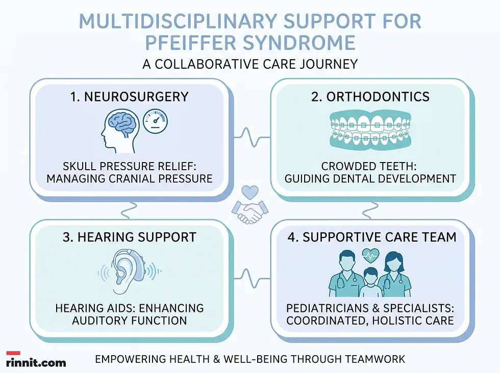
Treatment is multifaceted and typically involves a team of specialists, including neurosurgeons and plastic surgeons. The primary goal is to ensure the brain has enough space to grow and to prevent life-threatening complications. [1]
Pressure Relief: Shunts may be inserted to drain excess fluid (hydrocephalus) and alleviate intracranial pressure, usually before the infant is four months old.
Skull Expansion: Surgeries to open the fused sutures are often performed within the first year of life to allow for normal brain expansion.
Reconstructive Surgery: Later procedures focus on correcting facial asymmetry, improving breathing pathways, and addressing dental or hearing issues. [1]
Conclusion
Pfeiffer syndrome is a complex, lifelong condition that requires early diagnosis and a coordinated medical response. While the challenges can be significant, particularly for those with Type 2 or Type 3, modern surgical techniques have greatly improved the quality of life and outlook for affected children. With consistent medical follow-ups and specialized support, children with Pfeiffer syndrome can overcome many of the physical barriers associated with the condition.
Related Reading
Frequently Asked Questions
Is Pfeiffer syndrome a form of dwarfism?
No, Pfeiffer syndrome is primarily a disorder of bone fusion (craniosynostosis) and skeletal development in the hands and feet, rather than a condition affecting overall height in the way traditional dwarfism does.
Can Pfeiffer syndrome be detected before birth?
Yes, prenatal ultrasounds and MRIs can often identify the skeletal abnormalities associated with the condition during the second or third trimester.
What is a “cloverleaf” skull?
Found in Type 2 Pfeiffer syndrome, a cloverleaf skull occurs when multiple sutures fuse prematurely, causing the head to bulge at the top and sides in a shape resembling a three-lobed leaf.
Do children with Pfeiffer syndrome have intellectual disabilities?
Children with Type 1 typically have normal intelligence. Types 2 and 3 may involve intellectual disabilities, often depending on the severity of the brain compression and the timing of surgical intervention.
Is there a cure for the genetic mutation?
There is currently no cure for the underlying genetic mutation, but surgeries can effectively manage and correct the physical symptoms and complications it causes.
How many surgeries will a child need?
The number of surgeries varies widely based on the type and severity, but most children require multiple procedures throughout their early childhood and adolescence.
Does Pfeiffer syndrome affect hearing?
Yes, many children experience hearing loss due to the abnormal shape of the ear canals or the bones in the middle ear.
Is the condition painful for the baby?
The primary concern is the pressure inside the skull, which can cause discomfort. Surgeries are performed early to relieve this pressure and ensure the child’s comfort.
Can parents with the gene have a healthy child?
Yes. With an autosomal dominant condition, there is a 50% chance the child will inherit the gene and a 50% chance they will not.
How is Pfeiffer syndrome diagnosed after birth?
Doctors look at the shape of the head, face, hands, and feet, and use imaging such as X-rays or CT scans to see which skull sutures have closed. Genetic testing can confirm a change in an FGFR gene.
References
Cleveland Clinic. Pfeiffer Syndrome. Cleveland Clinic Health Library. Last updated July 12, 2023. https://my.clevelandclinic.org/health/diseases/25125-pfeiffer-syndrome
Way BLM, Khonsari RH, Karunakaran T, et al. Correcting exorbitism by monobloc frontofacial advancement in Crouzon-Pfeiffer syndrome: an age-specific, time-related, controlled study. Plast Reconstr Surg. 2019;143(1):121e-132e. PMID: 30589795. DOI: 10.1097/PRS.0000000000005105
Richardson IJ, Wager LE, Recker MJ, et al. Morbidity associated with anterior versus posterior cranial vault expansion for early treatment of syndromic craniosynostosis: a systematic review and meta-analysis. J Oral Maxillofac Surg. 2022;80(4):651-661. PMID: 34863645. DOI: 10.1016/j.joms.2021.10.022
Jayakumar NK, Pendem S, Vudatha RP, Muthu MS. Impact of Lefort III/monobloc advancement on midface growth in children with syndromic craniosynostosis: a systematic review. J Craniomaxillofac Surg. 2025;53(5):568-575. PMID: 39915153. DOI: 10.1016/j.jcms.2025.01.001
Medical disclaimer
This article is for informational purposes only and is not a substitute for professional medical advice, diagnosis, or treatment. Always seek the advice of your physician or other qualified health provider with any questions you may have about a medical condition.



Comments