Osteoid Osteoma: Symptoms, Causes, and Treatment Guide

Updated: 3 days ago
Medically reviewed by Dr. Baraa Alnahhal, MD · Last reviewed: October 2026
Osteoid osteoma is a small, benign bone tumor that usually affects the long bones of the legs and causes a dull, aching pain that gets worse at night. It is not cancer and may eventually go away on its own, but it can disrupt sleep and daily activity. Treatment ranges from over-the-counter pain relievers to minimally invasive ablation or surgery, and the long-term outlook is very good [1].
Quick Answer: What is osteoid osteoma?
Osteoid osteoma is a small, noncancerous bone tumor, usually less than one inch across, that most often develops in the shin or thigh bone of people between 5 and 25. Its hallmark is deep, aching pain that worsens at night and is often eased by anti-inflammatory medicine. Some resolve over years; others need radiofrequency ablation or surgical curettage. [1]
What the research says
Ablation works well across many patients. A systematic review of 36 studies covering 1,798 people and 1,863 percutaneous ablations found an overall success rate of 91.9%, with recurrence in 5.6% and complications in 2.5%; radiofrequency ablation and cryoablation performed similarly (Lindquester et al., Skeletal Radiol, 2020).
For spinal tumors, ablation matched surgery. A meta-analysis of 31 studies (749 patients) with spinal osteoid osteoma found mean success rates of 88.6% with radiofrequency ablation and 85.6% with surgical excision, with complications in 4.4% versus 7.8% (Sangiorgio et al., Eur Spine J, 2023).
What Is Osteoid Osteoma?
An osteoid osteoma is a small, benign bone tumor that does not spread to other parts of the body. These tumors usually measure less than 1 inch (2.5 centimeters) across. Even so, they can cause significant discomfort because of the way they interact with the surrounding bone and nerves [1].
They are most common in the long bones, especially the femur (thigh bone) and tibia (shin bone), but they can also develop in the arms, hands, fingers, feet, ankles, and spine. Osteoid osteomas make up roughly 10% of all benign bone tumors diagnosed today [1].
Feature | Osteoid osteoma | Osteoblastoma (for comparison) |
Tumor size | Small (usually under 1 inch) | Larger (over 2 cm) |
Common locations | Long bones (legs, arms) | Spine (about 40%), legs, feet |
Pain pattern | Worsens significantly at night | Not typically worse at night |
Recurrence risk | Low | Higher |
Cancer risk | Benign (noncancerous) | Benign (noncancerous) |
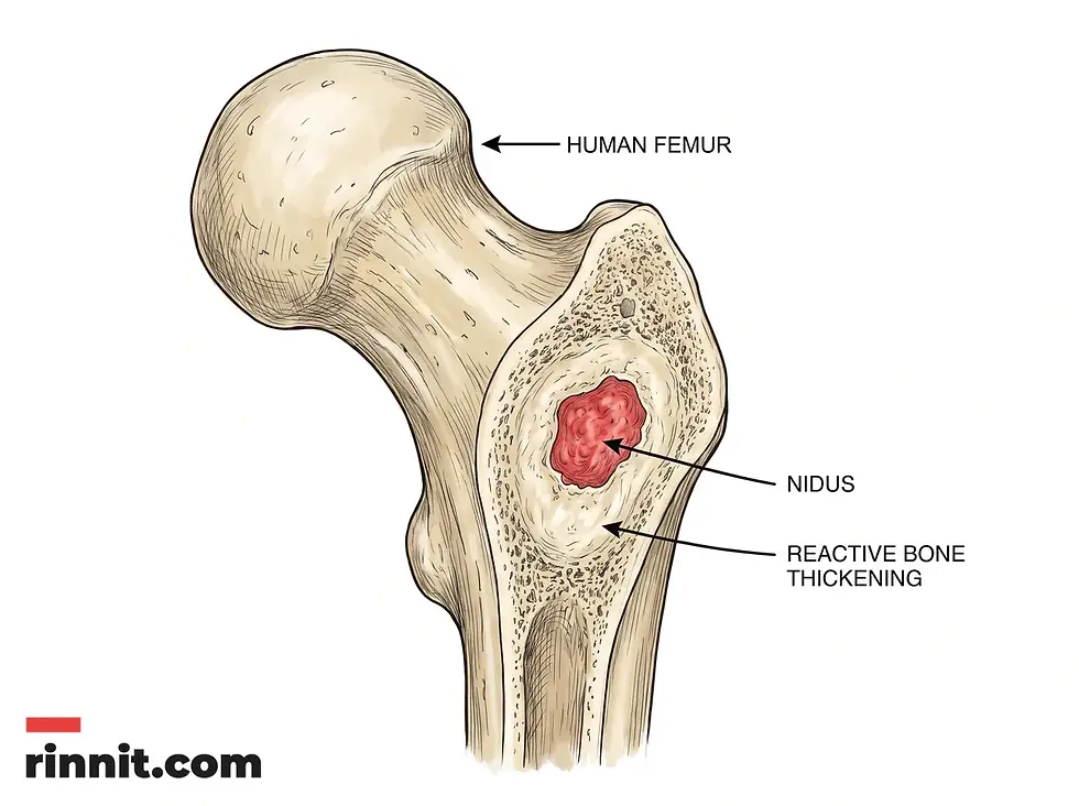
An osteoid osteoma in the femur: a small central core (the nidus) surrounded by thickened bone.
What are the symptoms and complications of osteoid osteoma?
The main symptom is a persistent, dull, aching pain. A distinctive feature is that the pain often gets worse at night and may wake you from sleep. Physical activity usually has little effect on how much it hurts [1].
If the tumor sits near a joint or in the spine, it can cause other symptoms and physical changes [1].
Possible physical symptoms:
Bone deformity: changes in the shape or structure of the affected bone [1].
Gait changes: a different walking pattern or a noticeable limp [1].
Joint problems: swelling (effusion), stiffness, or early osteoarthritis [1].
Muscle changes: a decrease in muscle size (atrophy) in the affected limb [1].
Structural effects: one leg looking longer than the other, or scoliosis or sciatica when the tumor is in the spine [1].
Symptom group | What you may notice |
Main pain | Deep, dull ache; worse at night; relieved by NSAIDs |
Structural changes | Muscle atrophy, leg length difference, bone deformity |
Joint effects | Stiffness, swelling, contractures, osteoarthritis |
Spinal effects | Scoliosis (curvature), sciatica (nerve pain) |
What causes osteoid osteoma, and what are the risks?
Researchers have not found the exact cause of osteoid osteoma. These tumors may come from abnormal cell growth (neoplasia), local bone inflammation, or an earlier injury to the area [1].
What is well documented is who gets it. Osteoid osteomas mostly develop in children and young adults between 5 and 25, and men are diagnosed three times as often as women [1].

Who is most affected, and the typical symptoms, including the night-time pain pattern.
How is osteoid osteoma diagnosed?
Diagnosis begins with a physical exam and a detailed review of your pain pattern. Your provider will ask how long and how severe the pain has been and whether over-the-counter pain relievers help [1].
X-ray: often the first test. The tumor typically shows as a patch of thickened bone around a small, clear core called the nidus [1].
Three-phase bone scan: a radioactive tracer is injected into the bloodstream to pinpoint the tumor’s exact location [1].
CT scan: gives a highly detailed image of the bone and is considered the most effective way to see the tumor’s structure [1].
MRI: less accurate for finding the tumor itself, but often used to rule out more serious conditions such as bone cancer [1].
Biopsy: sometimes a small tissue sample is examined under a microscope to confirm the diagnosis and rule out other bone disorders [1].
How is osteoid osteoma managed and treated?
An osteoid osteoma may resolve on its own without treatment, although this can take several years. If you have significant pain or limits on activity, several treatment paths are available [1].
Conservative Management
Nonsteroidal anti-inflammatory drugs (NSAIDs) such as aspirin, ibuprofen, or naproxen are the first line of treatment. They ease the characteristic night pain and may also help the tumor shrink over time. On average, symptoms managed this way resolve within about 33 months [1].
Minimally Invasive and Surgical Procedures
CT-guided radiofrequency ablation (RFA): a minimally invasive procedure in which a CT scan is used to find the tumor and a probe is inserted to destroy the tumor cells with heat. It has about a 90% success rate and allows a quick return to normal activity [1].
Curettage: a traditional operation in which the surgeon makes an incision to reach the bone and scrapes out the whole tumor [1].
Treatment | Benefits | Things to consider |
NSAIDs | Noninvasive, effective pain relief | It can take years for the tumor to resolve |
Radiofrequency ablation | About 90% success, outpatient procedure, fast recovery | Small risk of skin burns or nerve irritation |
Surgical curettage | Immediate, complete removal of the tumor | Needs anesthesia and a longer recovery |

Choosing a path after diagnosis: observation, NSAID medicine, or intervention with ablation or curettage.
What is the outlook and long-term recovery for osteoid osteoma?
The outlook is excellent. Whether the tumor resolves on its own, with medicine, or after a procedure, most people go on to live full, active lives without long-term problems [1].
Recovery time depends on the treatment. After radiofrequency ablation, many people return to school or work within two to three days. Traditional surgery may mean a short hospital stay and several weeks of physical therapy. After any procedure, you are usually advised to limit strenuous exercise for a few months so the bone can regain its full strength [1].
What is the bottom line on osteoid osteoma?
If you or your child has persistent bone pain that gets worse at night, get a medical evaluation. Osteoid osteoma is benign, but a professional diagnosis is essential to rule out other conditions and plan effective pain relief. Early treatment can restore sleep and help prevent structural bone changes.
Frequently Asked Questions
Is an osteoid osteoma cancerous?
No. It is a benign (noncancerous) tumor. It does not spread to other parts of the body and is not life-threatening.
Why does the pain get worse at night?
The exact reason is not fully understood, but pain that worsens at night is a hallmark of this condition. It often improves with anti-inflammatory medicine taken before bed.
Can it go away without surgery?
Yes. Some osteoid osteomas resolve on their own over several years. Many people still choose treatment to end persistent pain and sleep disruption.
What is the "nidus" in an osteoid osteoma?
The nidus is the small central core of the tumor where the abnormal cell growth occurs. A zone of dense, reactive bone usually surrounds it.
Is radiofrequency ablation painful?
The procedure is done under anesthesia (general or regional), so you do not feel pain during treatment. Most people get significant pain relief within days.
Who is most likely to get this tumor?
Osteoid osteoma is most common in children and young adults between 5 and 25, and it affects males about three times more often than females.
Will I need physical therapy after surgery?
If you have traditional surgical curettage, physical therapy is often recommended to help you regain strength and mobility in the affected limb.
Can an osteoid osteoma grow back?
Recurrence is uncommon, especially after successful surgical removal or radiofrequency ablation. If the pain returns, a follow-up evaluation is needed.
Are there long-term effects on bone strength?
Once the tumor is removed or resolves, the bone typically heals and regains its full strength. Temporary activity limits are usually advised while it heals.
Related Resources
Related reading on Rinnit:
References
Cleveland Clinic. Osteoid osteoma. Cleveland Clinic Health Library. Medically reviewed; last updated February 4, 2022. https://my.clevelandclinic.org/health/diseases/22405-osteoid-osteoma
Lindquester WS, Crowley J, Hawkins CM. Percutaneous thermal ablation for treatment of osteoid osteoma: a systematic review and analysis. Skeletal Radiol. 2020;49(9):1403-1411. PMID: 32270226. DOI: 10.1007/s00256-020-03435-7
Sangiorgio A, Oldrini LM, Candrian C, et al. Radiofrequency ablation is as safe and effective as surgical excision for spinal osteoid osteoma: a systematic review and meta-analysis. Eur Spine J. 2023;32(1):210-220 (published online 2022). PMID: 36214895. DOI: 10.1007/s00586-022-07411-8
Medical disclaimer
This article is for informational purposes only and is not a substitute for professional medical advice, diagnosis, or treatment. Always seek the advice of your physician or other qualified health provider with any questions you may have about a medical condition.



Comments