Ossifying Fibroma: Symptoms, Causes, Diagnosis, Treatment, and Outlook — What You Need to Know

Updated: 2 days ago
Medically reviewed by Dr. Baraa Alnahhal, MD · Last reviewed: September 2026
TL;DR: The Essentials
Ossifying fibroma is a rare, noncancerous (benign) tumor that primarily develops in the jawbones or other facial bones. It is classified as a fibro-osseous lesion, meaning healthy bone is replaced by fibrous tissue containing bone-like material. While often asymptomatic and slow-growing, these tumors require surgical removal because they can destroy healthy bone, cause teeth to shift, and lead to facial fractures. Recurrence is common, particularly in children, necessitating long-term follow-up care.
Quick Answer: What Is Ossifying Fibroma?
An ossifying fibroma is a rare, benign bone tumor that most commonly affects the mandible (lower jaw) and maxilla (upper jaw). The term "ossifying" refers to the process where fibrous connective tissue hardens into bone or bone-like material where it does not belong. These tumors are slow-growing but persistent, often replacing healthy bone structure with abnormal fibrous tissue.
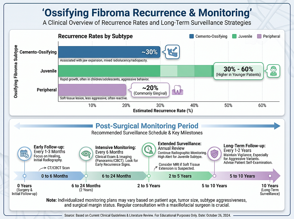
There are three primary types: cemento-ossifying fibroma (affecting the tooth roots), juvenile ossifying fibroma (an aggressive form seen in children), and peripheral ossifying fibroma (occurring on the gums). Because they can cause significant dental displacement and weaken the facial skeleton, surgical removal by an oral and maxillofacial surgeon is the standard treatment. Unlike some other benign growths, ossifying fibromas do not resolve on their own and have a notable tendency to grow back, requiring up to 10 years of clinical monitoring after surgery.
Understanding the Types of Ossifying Fibroma
Ossifying fibromas are categorized based on their location, growth rate, and the age group they typically affect. Understanding these distinctions is critical for determining the appropriate surgical approach and follow-up schedule.
Type | Primary Location | Typical Age Group | Growth Characteristics |
|---|---|---|---|
Cemento-Ossifying Fibroma | Mandible (Lower Jaw) | Adults (30s–40s), primarily females | Slow-growing; forms near tooth cementum |
Juvenile Ossifying Fibroma | Any part of the skull | Children (Ages 5–15) | Aggressive and fast-growing |
Peripheral Ossifying Fibroma | Maxilla (Upper Jaw) gums | Adolescents and young adults | Develops between teeth on the gingiva |

Symptoms and Warning Signs
Most individuals with an ossifying fibroma do not experience pain, and the tumor is often discovered incidentally during routine dental X-rays. However, as the tumor expands, it can cause visible changes and functional issues.
Common Clinical Presentations
Facial Swelling: Visible edema or asymmetry in the jaw or face.
Dental Shifting: Teeth may move, become loose, or shift out of alignment as the tumor displaces bone.
Nasal Congestion: Tumors in the upper jaw or skull can block nasal passages.
Bulging Eyes: Aggressive juvenile forms may affect the eye sockets (orbits).
Gum Abnormalities: Red bumps or swollen areas on the gums, particularly in peripheral types.
Symptom | Associated Type | Severity Level |
|---|---|---|
Asymptomatic (No symptoms) | All Types | Common in early stages |
Shifting or loose teeth | Cemento-ossifying | Moderate to High |
Bulging eyes (Proptosis) | Juvenile | High (Aggressive) |
Red gum bumps/ulcers | Peripheral | Moderate |
Causes and Risk Factors
The exact cause of ossifying fibroma remains unknown to medical experts. However, several factors are believed to potentially trigger the abnormal growth of fibrous tissue in the bone.
Potential Triggers
Trauma: Previous injury to the face or jawbones.
Dental Plaque: Significant buildup of plaque or chronic gum irritation.
Dental Procedures: Complications or trauma resulting from past oral surgeries.
Genetics: Particularly in juvenile forms, though specific markers are still being studied.

Diagnosis and Professional Testing
Because ossifying fibromas are rare and often painless, professional imaging is required to identify the lesion and differentiate it from other bone disorders.
Dental X-rays: Usually the first tool to detect abnormal bone density.
CT Scan: Provides detailed 3D views of the tumor's size and its impact on surrounding bone.
MRI: Useful for assessing the involvement of soft tissues and connective structures.
Biopsy: The definitive diagnostic tool. A tissue sample is examined in a lab, typically after the tumor has been surgically removed, to confirm it is benign.
Treatment and Management Strategies
Surgical intervention is the only effective treatment for ossifying fibroma. Unlike non-ossifying fibromas, these lesions will not disappear on their own and will continue to grow until removed.
Surgical Intervention
An oral and maxillofacial surgeon typically performs the procedure. The goal is complete removal of the tumor to minimize the risk of it growing back. If the tumor has destroyed a large portion of the jawbone, a bone graft may be necessary to restore the structural integrity of the face and support future dental implants or reconstructions.
Complications of Untreated Tumors
Complication | Impact on Health |
|---|---|
Facial Fracture | Weakened bone is highly susceptible to breaks from minor impact. |
Root Reabsorption | The tumor causes the body to dissolve tooth roots, leading to permanent tooth loss. |
Structural Deformity | Large tumors can permanently alter facial symmetry and appearance. |
Outlook and Recurrence Rates
The prognosis for ossifying fibroma is generally good because the tumors are noncancerous. However, the high rate of recurrence makes long-term monitoring essential. Children are significantly more likely to experience regrowth than adults.
Type | Recurrence Rate | Monitoring Requirement |
|---|---|---|
Cemento-Ossifying | ~30% | Annual X-rays for 10 years |
Juvenile | 30% – 60% | Frequent imaging; aggressive follow-up |
Peripheral | ~20% | Regular dental check-ups |
Frequently Asked Questions
Can an ossifying fibroma turn into cancer?
No. There is no documented evidence of an ossifying fibroma becoming malignant. They are strictly benign tumors.
What is the difference between ossifying and non-ossifying fibromas?
Ossifying fibromas harden into bone-like material and usually affect the face and jaw. Non-ossifying fibromas do not harden, typically affect leg bones in children, and often disappear on their own.
Is surgery always necessary?
Yes. Because these tumors destroy healthy bone and do not stop growing, surgical removal is required to prevent fractures and tooth loss.
How long does the surgery take?
The duration depends on the tumor's size and location, but most procedures are completed within a few hours by an oral surgeon.
Will I need a bone graft?
If the tumor is large and has replaced a significant amount of healthy jawbone, a graft will be used to fill the void and strengthen the jaw.
Why do these tumors grow back so often?
Their persistent nature and the difficulty of removing every microscopic trace of the fibrous tissue contribute to the high recurrence rates.
Can children get this?
Yes. The "juvenile" type specifically affects children between ages 5 and 15 and is more aggressive than the adult form.
Does insurance cover the removal?
Most medical and dental insurance plans cover the removal of benign tumors, though you should verify with your provider regarding bone grafts.
How often will I need follow-up X-rays?
Standard protocol involves regular imaging for up to 10 years after surgery to ensure early detection of any recurrence.
Can dental plaque really cause a bone tumor?
While not a direct cause, chronic irritation from plaque and gum disease is listed as a potential trigger for peripheral types.
Will I lose my teeth?
If the tumor is near the tooth roots, those teeth may shift or require removal. Bone grafting can help prepare the area for future replacements.
Is the surgery painful?
The procedure is performed under anesthesia. Post-operative pain is managed with medication, and most patients recover within a few weeks.
What happens if I ignore it?
The tumor will continue to expand, eventually leading to a broken jaw (facial fracture) or extensive dental damage.
Are there non-surgical treatments like radiation?
No. Radiation is not used for these benign tumors, as surgical excision is the gold standard and most effective method.
Who treats this condition?
You will typically work with a dentist for initial detection and an oral and maxillofacial surgeon for treatment and long-term care.
Conclusion and Next Steps
If you or your child experiences unexplained facial swelling, shifting teeth, or red bumps on the gums, consult a dental professional immediately. Early detection via X-ray can simplify the surgical process and protect the integrity of your jawbone. While the high recurrence rate requires a decade of vigilance, modern oral surgery effectively manages the condition and prevents long-term deformity.
Additional Resources
Visit the American Association of Oral and Maxillofacial Surgeons for patient resources on jaw tumors.
References
Cleveland Clinic. "Ossifying Fibroma." Medically Reviewed 10/10/2022. https://my.clevelandclinic.org/health/diseases/24275-ossifying-fibroma
Medical Disclaimer: This information is for educational purposes only and does not constitute medical advice. Always seek the advice of a qualified healthcare provider with any questions regarding a medical condition.
Comments