Optic Chiasm: Anatomy, Function, and Visual Health

Updated: 3 days ago
Medically reviewed by Dr. Baraa Alnahhal, MD · Last reviewed: October 2026
The optic chiasm is the X-shaped structure under the brain where the optic nerves from both eyes meet and partly cross. It helps you see one clear image and perceive depth, and because of its location it is at risk of damage that can change your vision [1].
Quick Answer: What is the optic chiasm?
The optic chiasm is where the optic nerves from both eyes meet and partly cross at the base of the brain, with only the inner-half fibers crossing. This helps the brain combine both eyes' views and judge depth. Nearby tumors and aneurysms can compress it, causing blind spots and loss of side vision in both eyes. Report any vision change. [1]
What the research says
Surgery relieving pressure from pituitary tumors usually improves vision. A meta-analysis of 35 case series of endoscopic endonasal surgery for pituitary adenomas found visual field deficits improved in 80.8% of patients and fully recovered in 40.4%, while 2.3% got worse (Muskens et al., Pituitary, 2017).
Vision can recover in pituitary apoplexy with or without surgery. A meta-analysis of 14 studies (457 cases) found 37% of patients had a visual acuity or field deficit at first examination, and the chance of visual improvement did not differ significantly between surgical and conservative treatment; the authors said the choice depends on the severity of initial deficits (Goshtasbi et al., World Neurosurg, 2019).
What Is the Optic Chiasm?
The optic nerve is the cable that connects your eye to your brain, and both optic nerves meet and cross at the chiasm. This intersection helps your brain combine what both eyes see, so when you open your eyes you get a single image. The chiasm is surrounded by blood vessels and glands [1].
Where Is the Optic Chiasm and What Does It Look Like?
The chiasm is at the bottom of the brain, below the hypothalamus and just above the pituitary gland, in an area called the suprasellar cistern. It is close to the third ventricle (fluid-filled cavities) and is surrounded by a loop of arteries called the circle of Willis [1]. It has an “X” shape, and “chiasm” comes from the Greek letter “chi” (X). On average it is about half an inch, or 13.5 millimeters, wide [1].

The optic chiasm sits between the hypothalamus above and the pituitary gland below.
What are the regions of the optic chiasm?
The optic chiasm is where nerve fibers cross, and this crossing helps the brain understand where things are in the left and right visual fields.
Region | Role |
Optic nerves | Carry visual messages from each eye; each contains nasal fibers (inner half of the retina, nearest the nose) and temporal fibers (outer half, farthest from the nose) |
Optic chiasm | Where some fibers cross to the other side of the brain: only the nasal fibers cross, and the temporal fibers stay on the same side |
Optic tracts | Continue after the crossing; each carries information from both eyes for one side of vision (the right tract carries what you see in your left visual field) |
This crossing helps your brain understand where things are in your left and right visual fields [1].
What does the optic chiasm do?
The optic chiasm lets the brain combine the views from both eyes into one image and supports depth perception, helping you see in three dimensions.
Creates a unified field of vision: each eye has a slightly different perspective, and the brain combines them so you see one image instead of two.
Enables depth perception (binocular vision): because the views from both eyes overlap, the brain picks up small differences between the two images, such as distance and depth, helping you see in three dimensions.
Source: Cleveland Clinic [1].
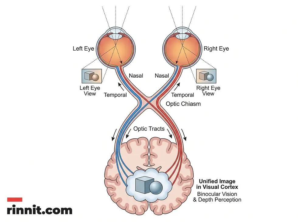
How crossing nerve fibers help the brain build one three-dimensional image from two eyes.
Which conditions can affect the optic chiasm?
Because of its location, the chiasm is at risk of damage from surrounding glands or blood vessels, which can lead to vision changes or even vision loss [1]. Conditions that can press on it include:
Tumors: craniopharyngioma, meningioma, optic nerve glioma, pituitary adenoma, and pilocytic astrocytoma
An aneurysm (a bulge or weak spot in an artery or blood vessel)
Pituitary apoplexy (sudden bleeding or loss of blood flow to the pituitary gland)
Other conditions can affect how the chiasm works [1]:
Condition | How it affects the chiasm |
Autoimmune conditions (such as neurosarcoidosis) | The immune system causes inflammation around the chiasm |
Demyelinating diseases (such as optic neuritis or multiple sclerosis) | Damage to the protective cover around nerves disrupts signals needed for vision |
Non-decussating retinal fugal fiber syndrome (achiasmatic syndrome) | A very rare syndrome in which nerve fibers do not cross at the chiasm |
Ocular albinism | A genetic condition that reduces pigmentation of eye structures and causes visual pathways to develop abnormally |
What Happens If the Optic Chiasm Is Damaged?
Damage may lead to [1]:
Blind spots
Blurred vision
Difficulty seeing colors
Loss of the outer sides of your vision in both eyes
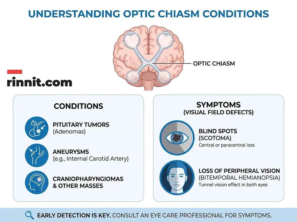
Conditions that can press on the optic chiasm and the visual field changes they can cause.
How can you protect your vision and optic chiasm?
Protecting the optic chiasm means telling your provider about any vision change and not waiting on new blind spots, blurred vision, color trouble or loss of side vision.
Let your provider know if anything about your vision seems off; they can check that your optic chiasm and its connected parts are working as they should.
Do not wait on new blind spots, blurred vision, trouble seeing colors, or loss of side vision, since these can reflect pressure on the chiasm. Call 911 or go to the emergency room for sudden vision loss, especially with a sudden severe headache, which can signal pituitary apoplexy.
What is the bottom line on the optic chiasm?
The optic chiasm is how you see one world instead of two. Its position near glands and blood vessels makes it vulnerable, so any change in vision deserves a prompt check by a healthcare provider [1].
Frequently Asked Questions
What is the optic chiasm?
The optic chiasm is the X-shaped structure at the base of the brain where the optic nerves from each eye meet and cross. It transmits visual information and helps your brain combine what both eyes see into a single image.
Where is the optic chiasm located?
The optic chiasm sits at the bottom of the brain, below the hypothalamus and just above the pituitary gland, in an area called the suprasellar cistern, close to the third ventricle and surrounded by the circle of Willis.
What does the optic chiasm do?
The optic chiasm helps create a unified field of vision, so you see one image instead of two, and enables depth perception (binocular vision), because the overlapping views from both eyes let the brain pick up differences in distance and depth.
Why is it called a chiasm?
“Chiasm” comes from the Greek letter chi (X), which matches the structure's X shape, formed where optic nerve fibers from both eyes cross.
Which fibers cross at the optic chiasm?
Only the nasal fibers, from the inner half of each eye closest to the nose, cross to the other side of the brain. The temporal fibers, from the outer half of each eye, stay on the same side.
What are the symptoms of optic chiasm damage?
Damage may cause blind spots, blurred vision, difficulty seeing colors, and loss of the outer sides of your vision in both eyes.
What conditions can affect the optic chiasm?
Tumors (craniopharyngioma, meningioma, optic nerve glioma, pituitary adenoma, pilocytic astrocytoma), an aneurysm, pituitary apoplexy, autoimmune conditions such as neurosarcoidosis, demyelinating diseases such as optic neuritis or multiple sclerosis, achiasmatic syndrome, and ocular albinism.
How does the optic chiasm help with depth perception?
Because the fields of view from both eyes overlap, your brain can pick up on small differences between the two images, such as distance and depth, so you see in three dimensions and judge how far away things are.
What are optic tracts?
After the fibers cross (or do not cross) at the chiasm, they continue as optic tracts. Each tract carries information from both eyes for one side of your vision; for example, the right optic tract carries what you see in your left visual field.
What should I do if my vision seems off?
Tell your healthcare provider. They can check that your optic chiasm and its connected parts are working as they should, and they can look for conditions pressing on it.
Related Resources
Related reading on Rinnit:
External resource for deeper reading:
References
Cleveland Clinic. Optic chiasm. Cleveland Clinic Health Library. Medically reviewed; last updated September 15, 2025. https://my.clevelandclinic.org/health/body/optic-chiasm
Muskens IS, Zamanipoor Najafabadi AH, Briceno V, et al. Visual outcomes after endoscopic endonasal pituitary adenoma resection: a systematic review and meta-analysis. Pituitary. 2017;20(5):539-552. PMID: 28643208. DOI: 10.1007/s11102-017-0815-9
Goshtasbi K, Abiri A, Sahyouni R, et al. Visual and endocrine recovery following conservative and surgical treatment of pituitary apoplexy: a meta-analysis. World Neurosurg. 2019;132:33-40. PMID: 31470146. DOI: 10.1016/j.wneu.2019.08.115
Medical disclaimer
This article is for informational purposes only and is not a substitute for professional medical advice, diagnosis, or treatment. Always seek the advice of your physician or other qualified health provider with any questions you may have about a medical condition.



Comments