Leptomeningeal Disease: Symptoms, Causes, and Management

Updated: 3 days ago
Medically reviewed by Dr. Baraa Alnahhal, MD · Last reviewed: October 2026
Leptomeningeal disease (LMD) is a serious complication in which cancer spreads to the cerebrospinal fluid and the membranes surrounding the brain and spinal cord. While currently incurable, healthcare providers focus on management strategies to slow the spread of cancer and ease neurological symptoms. Early detection through MRI and spinal tap allows interventions such as radiation, chemotherapy, and targeted therapies to help maintain comfort and quality of life [1].
Quick Answer: What is leptomeningeal disease?
Leptomeningeal disease (LMD) is an advanced cancer stage in which cancer cells, most often from breast, lung or melanoma, spread to the cerebrospinal fluid and membranes around the brain and spinal cord. Symptoms include headaches, confusion, vision problems and seizures. MRI and a spinal tap diagnose it. It is not curable, but radiation and chemotherapy can slow it. [1]
What the research says
Proton craniospinal irradiation delayed central nervous system progression compared with standard radiation. In the final analysis of a randomized phase 2 trial in non-small cell lung and breast cancer with leptomeningeal metastasis (42 patients assigned to proton craniospinal irradiation and 21 to photon involved-field radiotherapy), median CNS progression-free survival was 8.2 vs 2.3 months (P<.001) and median overall survival was 11.3 vs 4.9 months (P=.04); the authors suggest it should be considered when available (Yang et al., JAMA Oncol, 2025 [2]).
The same trial's earlier report found no added serious toxicity and explored other tumor types. Rates of grade 3 and 4 treatment-related adverse events did not differ between groups (P=.19), and in an exploratory group of 35 patients with other solid tumors who received proton craniospinal irradiation, median CNS progression-free survival was 5.8 months (Yang et al., J Clin Oncol, 2022 [3]).
What is Leptomeningeal Disease?
Leptomeningeal disease (LMD) is a major medical complication that occurs when cancer cells migrate from a primary tumor site to the cerebrospinal fluid (CSF) and the leptomeninges [1]. The leptomeninges are the delicate inner layers of the membranes that cover and protect the brain and spinal cord. This condition, sometimes referred to as leptomeningeal carcinomatosis or leptomeningeal metastases (LMM), typically appears in the later, more advanced stages of cancer.
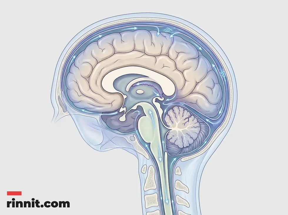
Cerebrospinal fluid circulates around the brain and spinal cord inside the leptomeninges, the protective membranes affected in leptomeningeal disease.
Approximately 5% of people diagnosed with metastatic cancer will develop LMD. While the condition is relatively rare, healthcare providers are observing an increase in cases as modern medical advancements allow people with cancer to live longer. Although there is currently no cure for LMD, the focus of medical care is to prevent further spread, manage symptoms, and preserve the patient's quality of life for as long as possible [1].
What are the neurological symptoms of leptomeningeal disease?
The presence of cancer cells in the cerebrospinal fluid can lead to a wide array of nervous system symptoms [1]. Because the CSF is in direct contact with the brain and spinal cord, the impact is often widespread.
Symptom Category | Common Manifestations |
|---|---|
Cranial Impacts | Persistent headaches, double vision, blurry vision, and hearing loss. |
Systemic Neurologic | Nausea, vomiting, confusion, and changes in alertness or mood. |
Physical Coordination | Weakness in the arms or legs, changes in gait (walking), and seizures. |
Localized Pain | Significant back and leg pain, along with numbness in the extremities. |
Many of these symptoms are worsened by hydrocephalus, a condition where the normal flow of cerebrospinal fluid is blocked, causing increased pressure within the skull. This often results in headaches that become significantly worse when a person is lying flat [1].
What causes leptomeningeal disease, and which cancers carry high risk?
Leptomeningeal disease is caused by the aggressive spread of cancer from another part of the body [1]. It is not a primary cancer but rather a metastatic development indicating that the underlying condition has become more serious.
While anyone with metastatic cancer can be affected, certain types of cancer carry a higher risk:
Breast and Lung Cancer: These are among the most common primary sources of LMD.
Melanoma: This aggressive skin cancer frequently spreads to the central nervous system.
Blood Cancers: Both acute lymphocytic leukemia and non-Hodgkin lymphoma are known risk factors for developing leptomeningeal complications.
How is leptomeningeal disease diagnosed and tested?
Accurately diagnosing LMD can be challenging, especially in cases where the spread of tumor cells is only microscopic [1]. Healthcare providers rely on a combination of advanced imaging and laboratory analysis.
The primary diagnostic tools include:
Magnetic Resonance Imaging (MRI): A detailed scan of both the brain and the spine is conducted to look for visible signs of tumor growth on the membranes.
Spinal Tap (Lumbar Puncture): A procedure to collect a sample of cerebrospinal fluid. This fluid is then tested in a laboratory to identify the presence of tumor cells or cancer cell DNA.

MRI of the brain and spine is a key diagnostic tool, used together with a spinal tap to test the cerebrospinal fluid.
How is leptomeningeal disease managed and treated?
The management of LMD is focused on slowing the progression of the cancer and ensuring the patient remains as comfortable as possible [1]. Treatment plans are highly individualized based on the patient's overall health and the type of primary cancer.
Primary Medical Treatments
Radiation therapy is frequently used to shrink tumors that are causing specific neurological symptoms, and in some cases, radiation is applied to the entire brain and spinal cord. Intrathecal chemotherapy involves delivering cancer-fighting drugs directly into the cerebrospinal fluid to target cells that systemic chemotherapy might not reach. Additionally, targeted therapies may be used to exploit specific genetic mutations within the cancer cells to stop their growth [1].

In intrathecal chemotherapy, cancer-fighting drugs are delivered directly into the cerebrospinal fluid.
Symptom Management and Comfort
To address the physical complications of LMD, providers may prescribe antiseizure medications or corticosteroids for pain relief. If hydrocephalus is present, a brain shunt may be surgically implanted to redirect excess fluid and reduce pressure. In the late stages of the disease, the focus may shift toward hospice care, prioritizing pain management and emotional support for both the patient and their loved ones [1].
What is the outlook for leptomeningeal disease?
Leptomeningeal disease is a major complication of advanced cancer. In general, people who receive treatment live for several months after diagnosis, while people who do not have treatment live for several weeks. These numbers are highly variable and depend on factors such as overall health and tumor type; new targeted therapies are helping people with some forms of LMD live longer than a year. Your healthcare team is the best resource for understanding what LMD means for your own situation [1].
What is the bottom line on leptomeningeal disease?
Leptomeningeal disease represents a significant challenge in the journey of advanced cancer care. While the diagnosis is serious and the condition is currently incurable, the evolution of targeted therapies and improved diagnostic techniques are providing new avenues for management. The primary goal of treatment remains the preservation of quality of life and the alleviation of symptoms through a multidisciplinary medical approach. Patients and their families are encouraged to work closely with their care teams to navigate the complexities of this diagnosis and access the necessary support resources [1].
Your next step: if you or a loved one is living with metastatic cancer and experiences new neurological symptoms such as persistent headaches or vision changes, contact your oncology team right away. Early intervention is key to managing the complications of LMD, and your team can also connect you with hospice and supportive care resources.
Frequently Asked Questions
What is leptomeningeal disease?
Leptomeningeal disease is a complication where cancer spreads to the cerebrospinal fluid and the membranes surrounding the brain and spinal cord.
Is LMD the same as brain metastases?
No. Brain metastases are tumors in the brain tissue itself, while LMD affects the fluid and membranes surrounding the brain. The two can occur separately or together.
What are the most common symptoms?
Common signs include headaches, nausea, confusion, weakness in the limbs, and vision changes.
Which cancers are most likely to cause LMD?
Breast cancer, lung cancer, melanoma, and certain types of leukemia and lymphoma carry the highest risk.
How is the condition diagnosed?
Diagnosis typically requires an MRI of the brain and spine and a spinal tap to analyze the cerebrospinal fluid.
Can leptomeningeal disease be cured?
Currently, there is no cure for LMD; treatment focuses on slowing the disease and managing symptoms.
What is hydrocephalus in the context of LMD?
Hydrocephalus in LMD is a buildup of pressure in the brain caused by cancer cells blocking the normal flow of cerebrospinal fluid.
What is intrathecal chemotherapy?
Intrathecal chemotherapy is a treatment where chemotherapy drugs are delivered directly into the cerebrospinal fluid.
How long can someone live with LMD?
Outlook varies. People who receive treatment generally live for several months after diagnosis, those without treatment for several weeks, and new targeted therapies are helping some people live longer than a year.
What is the role of hospice in LMD care?
Hospice focuses on providing comfort, pain relief, and emotional support during the final stages of the illness.
Related Reading
Brain Metastases (Secondary Brain Tumors): Symptoms, Causes, and Treatment covers cancer that spreads to the brain and how it is treated.
Lumbar Puncture (Spinal Tap): Purpose, Procedure, Recovery and Results explains how a spinal tap collects fluid and what recovery involves.
Cerebrospinal Fluid (CSF) Analysis: What It Tests, Procedure and Results explains what CSF tests look for and what results mean.
Cerebrospinal Fluid (CSF): Functions, Anatomy, and Clinical Importance explains what cerebrospinal fluid does around the brain and spine.
References
Cleveland Clinic. Leptomeningeal Disease. Cleveland Clinic. April 28, 2026. https://my.clevelandclinic.org/health/diseases/22737-leptomeningeal-disease
Yang JT, Yerramilli D, Pentsova E, et al. Proton craniospinal irradiation for patients with leptomeningeal metastasis: a randomized clinical trial. JAMA Oncol. 2025;11(11):1293-1301. PMID 40906462. https://doi.org/10.1001/jamaoncol.2025.3007
Yang JT, Wijetunga NA, Pentsova E, et al. Randomized phase II trial of proton craniospinal irradiation versus photon involved-field radiotherapy for patients with solid tumor leptomeningeal metastasis. J Clin Oncol. 2022;40(33):3858-3867. PMID 35802849. https://doi.org/10.1200/JCO.22.01148
Medical disclaimer
This article is for informational purposes only and is not a substitute for professional medical advice, diagnosis, or treatment. The clinical information in this guide reflects source material last reviewed in April 2026.



Comments