Fat Necrosis: Causes, Symptoms, and Treatment

Updated: 3 days ago
Medically reviewed by Dr. Baraa Alnahhal, MD · Last reviewed: October 17, 2026
Quick Answer: What is fat necrosis?
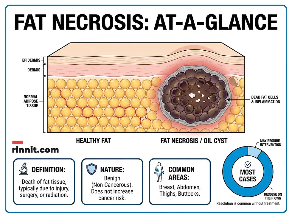
Fat necrosis is the localized death of fat tissue, most often in the breasts, abdomen or thighs after trauma, surgery or radiation. Disrupted blood supply leads to oil cysts that may calcify into hard nodules. These lumps can be alarming but are medically harmless and not linked to cancer. Most need only observation, though aspiration or excision are options.
What the research says
Fat necrosis after fat grafting to the breast is uncommon. A meta-analysis of 47 studies (4,425 participants) found a pooled fat necrosis incidence of 4.66% per participant (95% CI 1.88–8.35%), with wide variation between studies (Giannas et al., J Plast Reconstr Aesthet Surg, 2026 [2]).
Imaging changes after fat grafting rarely lead to treatment. In 22 studies of 2,073 women who had cosmetic breast augmentation with fat grafting, oil cysts appeared in 6.5%, calcifications in 4.5%, and fat necrosis in 1.2%. Palpable cysts occurred in 2.0%, and 67% of those were treated with aspiration (Ørholt et al., Plast Reconstr Surg, 2020 [3]).
Radiation can raise the risk. Across nine randomized trials involving 14,514 patients, partial breast irradiation was linked to higher odds of fat necrosis than whole breast irradiation (OR 1.72, P = 0.002) (Korzets et al., Breast Cancer Res Treat, 2019 [4]).
Understanding Fat Necrosis
Fat necrosis is a delayed biological response to adipose tissue injury. When fat cells are damaged by blunt force or surgical intervention, they lose their blood supply and begin to die. This process is slow and often progresses through several distinct stages before becoming noticeable [1].
Feature | Description |
Medical Category | Benign (non-cancerous) tissue death |
Primary Cause | Physical trauma or surgical injury |
Common Locations | Breasts, abdomen, buttocks, thighs |
Typical Outcome | Spontaneous resolution over time |
Contagious? | No |
Who is Affected?
While fat necrosis can affect anyone who sustains a significant injury to their fatty tissue, certain groups are at a higher risk due to anatomical factors or medical history [1].
High-Risk Group | Reason for Increased Risk |
People with Breasts | Breasts contain large, exposed areas of fatty tissue prone to trauma. |
Post-Surgical Patients | Procedures like breast reconstruction or fat grafting involve tissue manipulation. |
Radiation Recipients | Cancer treatments can damage the blood supply to local fat cells. |
Newborns | Traumatic delivery conditions can occasionally cause localized fat death. |
Symptoms and Clinical Appearance
The symptoms of fat necrosis vary depending on the severity of the initial injury and the stage of tissue repair. It is common for these changes to appear months or even years after the original event, with an average delay of 1.5 years [1].
Visual and Physical Changes
You may notice changes in the texture or appearance of the skin directly over the affected area [1]. In the breast, this can sometimes lead to structural changes that mimic more serious conditions [1].
Symptom Type | Common Observations |
Skin Texture | Dimpling, sagging, or thickening of the skin surface. |
Skin Color | Redness, bruising, or localized discoloration. |
Nipple Changes | Potential retraction (sinking in) of the nipple if necrosis is nearby. |
Internal Texture | Soft oil cysts in early stages; hard, calcified nodules later. |
Does It Hurt?
In most instances, fat necrosis is completely painless. However, some individuals report mild tenderness in the affected area [1]. A specific type known as subcutaneous fat necrosis, which is associated with panniculitis (inflammation of the fat layer), is more likely to cause chronic pain due to the underlying inflammatory response [1].
Causes and Triggering Events
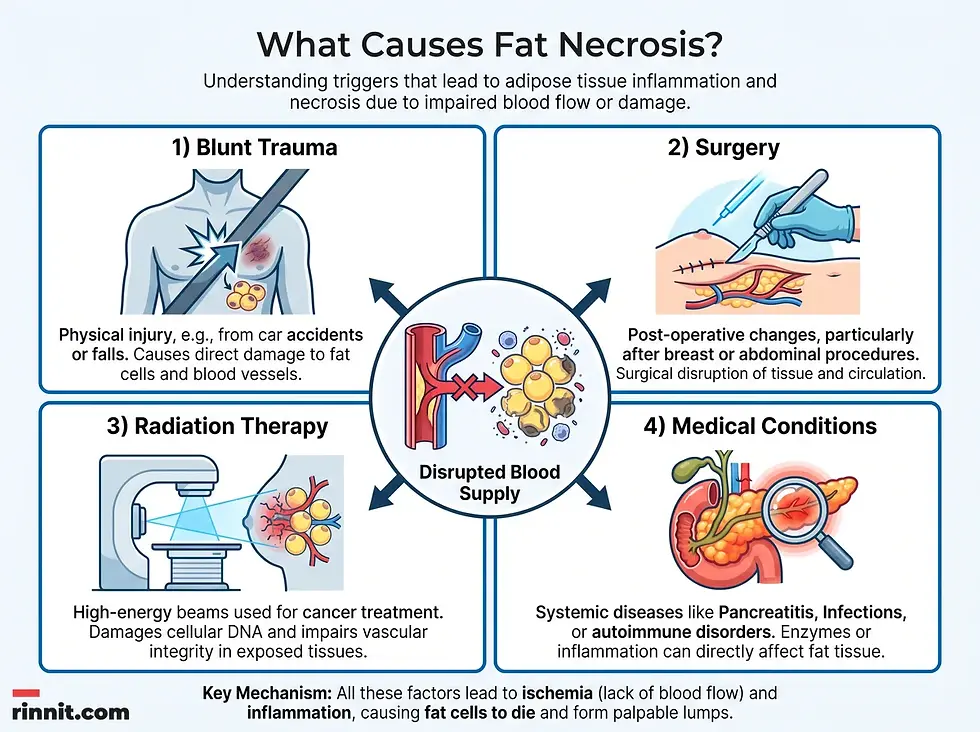
Fat necrosis occurs exclusively when the blood supply to fat cells is compromised, leading to cellular death. The triggers are almost always external or related to internal medical complications.
Trigger Category | Specific Examples |
Blunt Trauma | Car accidents (seatbelt impact), falls, or sports injuries. |
Therapeutic Surgery | Breast cancer surgery, mastectomies, or biopsies. |
Cosmetic Surgery | Tummy tucks, liposuction, or Brazilian butt lifts (BBL). |
Medical Treatments | Localized radiation therapy for cancer. |
Internal Disease | Acute pancreatitis (leakage of corrosive enzymes). |
The Role of Pancreatitis
In rare cases, severe acute pancreatitis can cause fat necrosis. When the pancreas is inflamed, it may leak corrosive enzymes into the abdominal cavity. These enzymes break down surrounding fat tissue, which can occasionally spread to the fat layer just beneath the skin (subcutaneous fat) in a condition called panniculitis [1].
Diagnosis and Rule-Out Testing
The primary challenge in diagnosing fat necrosis is its similarity to malignant tumors [1]. Because the lumps are often hard and may appear long after an injury is forgotten, healthcare providers must use precise testing to confirm the condition is benign [1].
Diagnostic Tools
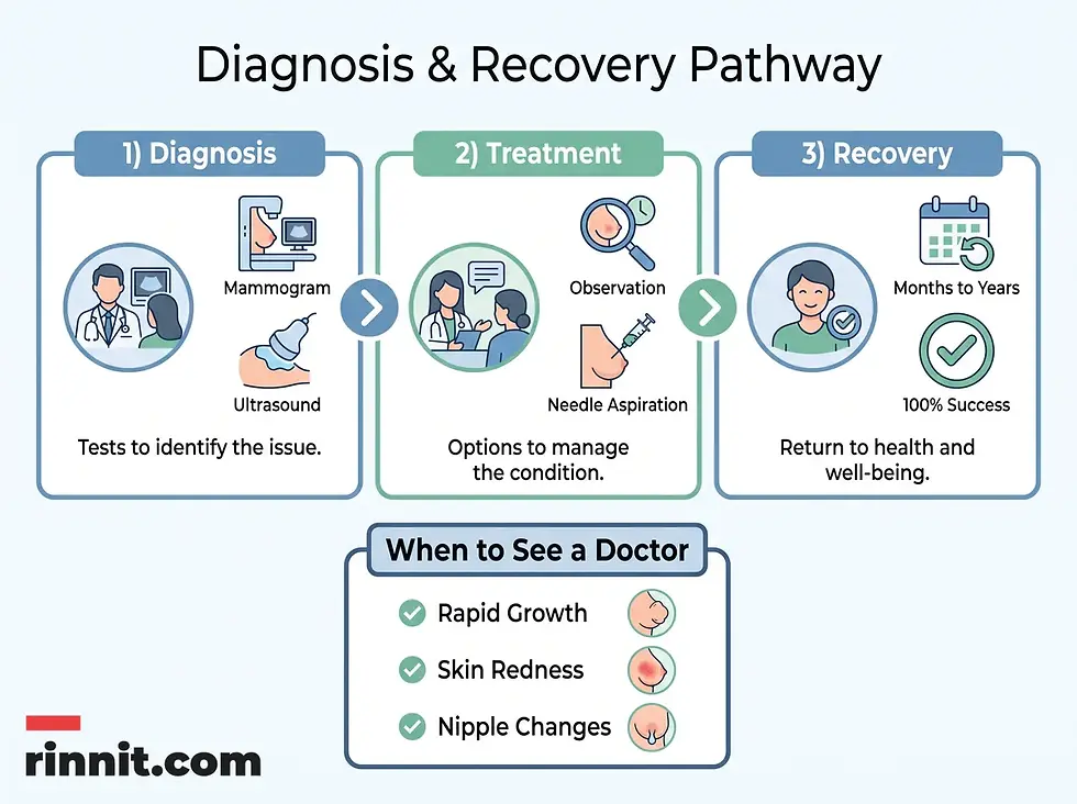
Radiology is the standard method for identifying fat necrosis, though a detailed health history is equally important for linking the lump to a past injury [1].
Test Type | Purpose in Fat Necrosis |
Detailed History | Linking the lump to surgeries or injuries from 1–2 years prior. |
Ultrasound | Visualizing oil cysts and distinguishing them from solid masses. |
Mammogram / X-ray | Identifying characteristic calcifications (hardened walls). |
MRI / CT Scan | Providing high-resolution views of tissue death and blood supply. |
Needle Biopsy | Extracting a tissue sample if cancer cannot be ruled out via imaging. |
Management and Treatment Options
For the vast majority of patients, fat necrosis requires no treatment. The body naturally breaks down the dead tissue and repairs the area over time [1]. However, if the lump causes significant anxiety, physical pain, or cosmetic concerns, several removal methods are available [1].
Method | How It Works |
Observation | "Watch and wait" approach; most lumps shrink on their own. |
Fine Needle Aspiration | A thin needle drains fluid from an oil cyst, causing it to collapse. |
Vacuum-Assisted Biopsy | A hollow needle suctions out small pieces of tissue under local anesthesia. |
Surgical Excision | Manual removal of the lump; reserved for large or suspicious masses. |
Risks of Surgical Removal
While surgery can remove a problematic lump, it is generally considered a last resort. Because fat necrosis is caused by tissue injury, the act of surgical cutting can potentially trigger new areas of necrosis in the surrounding fat [1].
Outlook and Recovery
The long-term outlook for fat necrosis is excellent. It is a self-limiting condition that does not lead to cancer or other life-threatening complications [1].
Spontaneous Resolution: Most lumps shrink and disappear without intervention [1].
Timelines: The full cycle of tissue breakdown and repair can take months to several years [1].
Post-Removal: If a lump is surgically removed, it may leave a small permanent scar or a slight indentation in the skin [1].
Fat Grafting: If necrosis occurs after a fat transfer (like a BBL), the procedure may be considered a failure, as the grafted tissue did not survive [1].
Important Note: While fat necrosis is harmless, any new or changing lump should be evaluated by a healthcare professional to ensure an accurate diagnosis and peace of mind.
Contact Your Provider If:
This article provides general information and is not a substitute for professional medical advice. Always consult a qualified healthcare provider about any new or changing lump.
Frequently Asked Questions
Can fat necrosis turn into cancer?
No, fat necrosis is a benign condition involving the death of fat cells. It does not contain cancerous cells and cannot transform into a malignancy. However, it can sometimes look like cancer on imaging tests, which is why doctors may perform a biopsy to be certain.
How long does it take for fat necrosis to go away?
The timeline varies significantly between individuals. It typically takes months for the body to begin breaking down the dead fat cells, and the entire process can last several years. During this time, the lump may seem to change in size or texture as it heals.
Is fat necrosis common after breast surgery?
Yes, it is a relatively common complication following breast procedures, including mastectomies, lumpectomies, and reconstructions. It occurs because these surgeries inevitably disrupt some of the local blood supply to the fatty tissue.
Does fat necrosis always cause a hard lump?
Not always. In its early stages, it may feel like a soft, fluid-filled pocket (an oil cyst). It only becomes a hard nodule if the walls of that cyst begin to calcify or harden over time.
Can you prevent fat necrosis?
Since it is caused by injury, there is no guaranteed way to prevent it other than avoiding trauma. In surgical settings, experienced surgeons use techniques to minimize tissue damage and preserve blood flow, but some degree of fat necrosis may still occur.
Is fat necrosis contagious?
No, fat necrosis is not an infection and cannot be spread from person to person. It is a localized reaction to physical injury or surgical trauma within an individual’s own body.
Why does fat necrosis happen after a car accident?
Blunt force trauma, such as the intense pressure from a seatbelt during a collision, can crush fat cells and sever the tiny blood vessels that supply them. This lack of oxygen and nutrients causes the fat tissue to die.
Can fat necrosis happen in the abdomen?
Yes, it frequently occurs in the abdominal area following procedures like tummy tucks (abdominoplasty) or after significant blunt trauma to the stomach region.
What is an oil cyst?
An oil cyst is a collection of liquid fat that leaks out of dying adipose cells. It is one of the early stages of fat necrosis. The body eventually reabsorbs this liquid, or the cyst may harden into a firm lump.
Does insurance cover fat necrosis removal?
Coverage depends on your specific insurance provider and the reason for removal. If the procedure is deemed medically necessary due to pain or infection, it is typically covered. If removed solely for cosmetic reasons, it may not be.
Additional Resources
For further reading, these authoritative external references support the information above:
American Cancer Society. Fat Necrosis and Oil Cysts in the Breast
Breast Cancer Now. Fat Necrosis Guide
StatPearls. Breast Fat Necrosis Pathophysiology
Content reviewed and produced following the Rinnit editorial framework. Last updated: October 8, 2026.
Related Reading
Mammogram: Types, How to Prepare, What Happens and Your Results explains the breast X-ray that often finds fat necrosis.
Breast Biopsy: Types, What to Expect, Recovery and Results describes how breast lumps are sampled to rule out cancer.
Breast Cysts: Symptoms, Causes, and Treatment covers fluid-filled breast lumps and how they differ from other lumps.
Understanding Panniculitis: Inflammation of Subcutaneous Fat explains inflammation of the fat layer under the skin.
References
Cleveland Clinic. Fat Necrosis. Cleveland Clinic. Medically reviewed; last updated September 12, 2022. https://my.clevelandclinic.org/health/diseases/24187-fat-necrosis
Giannas E, Fritsch AM, Jeon H, et al. Fat survival following autologous fat transfer to the native breast: a systematic review and meta-analysis. J Plast Reconstr Aesthet Surg. 2026;115:146-156. PMID 41791205. doi:10.1016/j.bjps.2026.02.017
Ørholt M, Larsen A, Hemmingsen MN, et al. Complications after breast augmentation with fat grafting: a systematic review. Plast Reconstr Surg. 2020;145(3):530e-537e. PMID 32097306. doi:10.1097/PRS.0000000000006569
Korzets Y, Fyles A, Shepshelovich D, et al. Toxicity and clinical outcomes of partial breast irradiation compared to whole breast irradiation for early-stage breast cancer: a systematic review and meta-analysis. Breast Cancer Res Treat. 2019;175(3):531-545. PMID 30929116. doi:10.1007/s10549-019-05209-9



Comments