Veins: Understanding Anatomy, Function, and the Venous System

Updated: 3 days ago
Medically reviewed by Dr. Baraa Alnahhal, MD · Last reviewed: October 17, 2026
Editorial note: This article is provided for educational purposes only. It is not medical advice. Always consult a qualified healthcare professional about your individual health situation.
Quick Answer
Veins are blood vessels that collect oxygen-poor blood from the body’s tissues and return it to the heart. They hold approximately 75% of the body’s total blood volume and are categorized into deep, superficial, and perforating types. While most veins carry oxygen-depleted blood, the pulmonary veins are a unique exception, transporting oxygen-rich blood from the lungs back to the heart.
What the research says
The calf muscle pump is often impaired in chronic venous disease, and exercise may help. A review of 10 studies found that people with chronic venous disease had a lower calf pump ejection fraction than healthy people (a 15.9% reduction, P < .001) and a higher venous filling index. Calf pump dysfunction was present in 55% of patients, though this did not reach significance on meta-analysis. Exercise programs in people with active and healed venous ulcers increased calf pump function but not valve competence. The authors say clinical trials are still needed (Williams et al., J Vasc Surg Venous Lymphat Disord, 2014 [1]).
Vein disease is very common, and several risk factors are known. A systematic review of 32 studies from 6 continents (19 of them pooled for prevalence) estimated that 19% of adults have varicose veins (CEAP class C2) and 8% have leg swelling (C3), with 4% having skin changes, 1% healed ulcers and 0.42% active ulcers. Reported risk factors included female sex (odds ratio 2.26, 95% CI 2.16–2.36), increasing age, obesity, prolonged standing, family history and having had children. The authors note significant variation between studies (Salim et al., Ann Surg, 2021 [2]).
Anatomical Structure of Veins
Veins are composed of three distinct tissue layers, similar to arteries but with thinner, less muscular walls due to the lower pressure environment of the venous system. The innermost layer, the tunica intima, is lined with smooth endothelial cells to facilitate easy blood movement. The middle layer, the tunica media, contains smooth muscle that allows the vessel to adjust its diameter. The outermost layer, the tunica adventitia, provides structural shape and integrity. A defining feature of many veins, particularly those in the legs, is the presence of one-way valves that ensure blood moves only toward the heart.
Layer | Scientific Name | Primary Function |
Inner Layer | Tunica Intima | Smooth lining for low-friction flow |
Middle Layer | Tunica Media | Muscle cells for dilation and constriction |
Outer Layer | Tunica Adventitia | Structural support and vessel shape |

The Venous Network and Circuits
The venous system is organized into two primary circuits: the systemic circuit and the pulmonary circuit. The systemic circuit spans the entire body, collecting oxygen-poor blood and waste products like carbon dioxide through a network that begins at the capillaries and venules. This blood eventually enters the heart through the superior and inferior vena cava. The pulmonary circuit is shorter, moving blood into the lungs for re-oxygenation before it returns to the heart via the pulmonary veins—the only instance where veins carry oxygen-rich blood.
Vein Type | Location | Key Characteristic |
Deep Veins | Inside muscles/along bones | Move 90% of blood back from legs |
Superficial Veins | Just beneath the skin | Visible and carry blood from outer tissues |
Perforating Veins | Connecting deep and superficial | Feature valves to prevent deep-to-surface backflow |
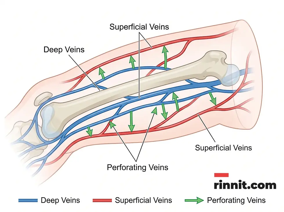
Mechanics of Blood Flow: The “Second Heart”
Because veins operate under low pressure, they require external forces to push blood against gravity, particularly from the lower limbs. Breathing creates a suction force in the chest that helps draw blood upward. However, the most significant force is the peripheral heart or “second heart,” which refers to the calf muscles. Each step taken squeezes the deep veins in the feet and calves, propelling blood toward the thighs and heart. This mechanism is dynamic, adjusting its pace to the level of physical activity, which is why walking and movement are critical for vascular health.

Common Vascular Considerations
When the one-way valves in the veins become damaged or weakened, blood can pool or flow backward, leading to several common conditions. Chronic venous insufficiency occurs when the venous walls or valves do not work effectively, making it difficult for blood to return to the heart from the legs. Other notable conditions include deep vein thrombosis (DVT), where a blood clot forms in a deep vein, and varicose veins, which are swollen, twisted veins visible just under the surface of the skin.
Conclusion
Veins are a resilient and sophisticated component of the circulatory system, responsible for the vital task of returning blood for re-oxygenation. Their unique anatomical features, such as one-way valves and thin-walled flexibility, allow them to manage the majority of the body’s blood volume efficiently. By understanding the importance of the calf muscle pump and the mechanics of venous return, individuals can better appreciate the role of movement in maintaining long-term vascular health.
Call to Action
Support your venous health by incorporating regular movement into your daily routine to keep your “second heart” active. If you experience persistent leg swelling, pain, or visible changes in your veins, consult a healthcare professional for a vascular evaluation. Staying active is the most effective way to ensure your circulatory system remains efficient.
Related reading: The Circulatory System: Your Body’s Life-Sustaining Transport Network | Sclerotherapy: A Minimally Invasive Treatment for Varicose and Spider Veins | Vein Ligation and Stripping for Varicose Veins
Sources: Williams et al., 2014 | Salim et al., 2021
Frequently Asked Questions
1. What is the main function of veins?
Veins' primary role is to return oxygen-poor blood from the body’s tissues back to the heart.
2. Do all veins carry oxygen-poor blood?
No, the pulmonary veins are the exception; they carry oxygen-rich blood from the lungs to the heart.
3. Why do veins look blue through the skin?
The blue appearance of veins is an optical illusion caused by how light is absorbed by the skin; blood in veins is actually dark red.
4. What are the three layers of a vein?
The three layers of a vein are the tunica intima (inner), tunica media (middle), and tunica adventitia (outer).
5. How do veins differ from arteries?
Veins have thinner walls, lower pressure, and often contain one-way valves, unlike arteries.
6. What is a venule?
A venule is a very small blood vessel that connects capillaries to larger veins.
7. What percentage of blood is held in the veins?
Nearly 75% of the body’s total blood volume is contained within the venous system.
8. What are deep veins?
Deep veins are veins located deep within the muscles and along bones that do the bulk of the work returning blood.
9. What is the largest vein in the body?
The great saphenous vein, a superficial vein in the leg, is the longest.
10. What is the “second heart”?
The “second heart” refers to the calf muscles, which pump blood upward when you walk or move.
11. What is the purpose of venous valves?
Venous valves are one-way flaps that prevent blood from flowing backward, especially in the legs.
12. What are perforating veins?
Perforating veins are short veins that connect the superficial venous system to the deep venous system.
13. How does breathing help blood flow?
The expansion of the lungs creates a suction effect that helps pull blood toward the heart.
14. What is deep vein thrombosis (DVT)?
Deep vein thrombosis (DVT) is a condition where a blood clot forms in one of the body’s deep veins, usually in the legs.
15. What causes varicose veins?
Varicose veins are often caused by weakened or damaged valves that allow blood to pool in the superficial veins.
References
Williams KJ, Ayekoloye O, Moore HM, Davies AH. The calf muscle pump revisited. J Vasc Surg Venous Lymphat Disord. 2014;2(3):329-334. PMID 26993396. https://doi.org/10.1016/j.jvsv.2013.10.053
Salim S, Machin M, Patterson BO, Onida S, Davies AH. Global Epidemiology of Chronic Venous Disease: A Systematic Review With Pooled Prevalence Analysis. Ann Surg. 2021;274(6):971-976. PMID 33214466. https://doi.org/10.1097/SLA.0000000000004631
Medical disclaimer
This content is for educational purposes and does not replace professional medical advice, diagnosis, or treatment.



Comments