Understanding the Uterus

Updated: 3 days ago
Medically reviewed by Dr. Baraa Alnahhal, MD · Last reviewed: October 2026
The uterus is a hollow, muscular organ central to the female reproductive system. It facilitates menstruation, provides the site for fertilized egg implantation, and supports fetal development during pregnancy. [1] Understanding its complex anatomy—including its layers, parts, and various physical positions—is essential for recognizing and managing common uterine conditions and maintaining overall reproductive well-being.
Quick Answer: What is the uterus?
The uterus is a pear-shaped, muscular organ in the pelvis between the bladder and rectum. [1] It sheds its lining during menstruation, hosts a fertilized egg for implantation and expands to nourish and protect a growing fetus during pregnancy. [1] Its muscles contract during labor to deliver the baby. [1] When not pregnant, it is roughly fist-sized and weighs about one ounce. [1]
What the research says
Low vitamin D is associated with fibroids. A systematic review of 69 studies found that 26 of 29 clinical studies examining hypovitaminosis D confirmed an association with fibroid prevalence, and a meta-analysis of 21 clinical studies showed women with fibroids had lower vitamin D concentrations than controls (mean difference -5.90 ng/mL, P < 0.001); the data are observational, so they show association rather than cause (Gutiérrez-Camacho et al., The Journal of Nutrition, 2026) [2].
Adenomyosis research is moving toward blood and urine tests. A systematic review of 27 studies found 39 distinct microRNAs significantly dysregulated in adenomyosis, with some studies showing diagnostic potential using serum, plasma or urine samples, but differences between studies precluded a meta-analysis and large validation studies are still needed (Buehler et al., Biomedicines, 2026) [3].
Pelvic floor training helps sexual function after hysterectomy. A meta-analysis of 6 randomized trials (776 women) found moderate-quality evidence that pelvic floor muscle training improved sexual function by 5 points (95% CI 4-6) on the Female Sexual Function Index compared with no intervention, with no or negligible effect on vaginal prolapse (risk difference 0; 95% CI -0.1 to 0.1) (de Oliveira et al., Brazilian Journal of Physical Therapy, 2024) [4].
Anatomy and Structure
The uterus is composed of several distinct parts and tissue layers, each serving a specific biological purpose. Its structure allows it to expand from the size of a lemon to that of a watermelon during pregnancy. [1]
The uterine wall consists of three specialized layers:

Functions in Reproduction
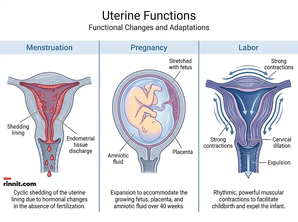
The uterus performs four primary roles that are critical to reproductive health. These functions are governed by hormonal changes and the muscular capability of the myometrium.
Menstrual Cycles: When pregnancy does not occur, the uterus sheds its endometrial lining, a process often accompanied by muscular contractions. [1]
Fertility: The uterus provides the necessary environment for a fertilized egg to implant and begin development. [1]
Pregnancy Support: The organ stretches to accommodate the fetus, while the placenta attaches to the uterine wall to deliver vital nutrients. [1]
Labor and Delivery: Regular contractions help open the cervix and push the baby through the birth canal during delivery. [1]
Common Uterine Positions and Conditions
While most uteruses tilt forward (anteverted), variations in position are common. For instance, approximately 20% to 25% of women have a retroverted uterus, which tilts backward toward the rectum. [1] Other variations include anteflexed, retroflexed, and midposition alignments. [1]
Various conditions can affect uterine health, leading to symptoms such as abnormal bleeding, pelvic pain, or infertility. [1]
Fibroids and Polyps: Noncancerous growths that can occur in the muscular wall or endometrial lining. [1]
Endometriosis and Adenomyosis: Conditions involving the abnormal growth of uterine-like tissue. [1]
Uterine Prolapse: A weakening of the pelvic floor muscles that causes the uterus to sag. [1]
Congenital Abnormalities: Structural differences present at birth, such as a heart-shaped (bicornuate) or divided (septate) uterus. [1]

Conclusion
As the central organ of the female reproductive system, the uterus is vital for fertility, pregnancy, and overall health. Its ability to transform and support life is one of the most remarkable features of human anatomy. By understanding its functions and staying aware of common symptoms, individuals can work with healthcare providers to ensure long-term reproductive wellness.
Frequently Asked Questions
What is the uterus?
The uterus is a hollow, pear-shaped muscular organ in the pelvis where a fetus develops during pregnancy.
How big is a non-pregnant uterus?
In its non-pregnant state, the uterus is about the size of a fist or a lemon, measuring roughly 3 inches long.
What are the three layers of the uterus?
The three layers are the perimetrium (outer), myometrium (muscular middle), and endometrium (inner lining).
What is the function of the endometrium?
The endometrium is the lining that thickens each month to prepare for pregnancy and sheds during menstruation if no pregnancy occurs.
What is a retroverted uterus?
A retroverted uterus is a common variation where the organ tilts backward toward the rectum instead of forward.
What are uterine fibroids?
Uterine fibroids are noncancerous tumors that grow in the muscular wall of the uterus.
How much does the uterus grow during pregnancy?
The uterus can stretch from the size of a lemon to the size of a watermelon, weighing up to 2 pounds by the end of pregnancy.
What is uterine prolapse?
Uterine prolapse occurs when pelvic floor muscles and ligaments weaken, causing the uterus to sag into the vaginal canal.
What symptoms suggest a uterine problem?
Symptoms include abnormal or heavy bleeding, severe pelvic pain, irregular periods, and pain during intercourse.
What is a bicornuate uterus?
A bicornuate uterus is a congenital abnormality where the organ is heart-shaped with two separate horns.
Related Reading
Understanding the Endometrium: The Essential Uterine Lining describes the uterine lining and its role in periods and pregnancy.
Endometriosis: Symptoms, Causes, and Treatment explains endometriosis pain, diagnosis and treatment options.
Adenomyosis: Complete Guide to Symptoms, Causes, Diagnosis & Treatment covers uterine lining tissue growing into the muscle wall.
Hysterectomy (Surgical Removal of the Uterus) explains types of hysterectomy, recovery and risks.
References
Cleveland Clinic. Uterus. Cleveland Clinic. Last updated March 18, 2026. https://my.clevelandclinic.org/health/body/22467-uterus
Gutiérrez-Camacho AI, Kim H, Prince L, et al. Vitamin D Deficiency Is Associated with Increased Uterine Fibroid Risk: A Systematic Review and Meta-analysis. The Journal of Nutrition. 2026:101838. PMID 42772682. https://doi.org/10.1016/j.tjnut.2026.101838
Buehler P, Vidal A, Vaineau C, et al. MicroRNAs as Biomarkers for Adenomyosis: A Systematic Review. Biomedicines. 2026;14(8):1764. PMID 42652148. https://doi.org/10.3390/biomedicines14081764
de Oliveira NFF, Santuzzi CH, da Conceição TV, et al. Pelvic floor muscle training for urinary symptoms, vaginal prolapse, sexual function, pelvic floor muscle strength, and quality of life after hysterectomy: a systematic review with meta-analyses. Brazilian Journal of Physical Therapy. 2024;28(5):101122. PMID 39426098. https://doi.org/10.1016/j.bjpt.2024.101122
Medical disclaimer
The information provided in this article is for educational purposes only and should not be used as a substitute for professional medical advice, diagnosis, or treatment. Always seek the advice of your physician or other qualified health provider with any questions you may have regarding a medical condition.



Comments