Understanding Retracted Eardrum: Symptoms, Stages, and Management

Updated: 3 days ago
Medically reviewed by Dr. Baraa Alnahhal, MD · Last reviewed: October 2026
A retracted eardrum occurs when an air pressure imbalance pulls the tympanic membrane inward toward the middle ear. Often caused by eustachian tube dysfunction, it can range from mild cases that resolve on their own to severe conditions requiring surgery to prevent permanent hearing loss.
Quick Answer
Retracted eardrum is a condition where the eardrum pulls toward the middle ear due to a vacuum effect caused by unequal air pressure. [1] Symptoms include earaches, a feeling of fullness, and temporary hearing loss. [1] While mild cases may resolve with monitoring or the Valsalva maneuver, severe retraction can lead to complications like cholesteatoma or bone damage, needing treatment or surgery. [1]
What the research says
Many deep retractions stay stable, but some progress. In a 10-year study of 64 children and 25 adults with asymptomatic deep pars tensa retraction, 19 children and 5 adults needed intervention and 5 children and 2 adults developed pars tensa cholesteatoma; the ears of 20 children, but none of the adults, returned to normal, and most adult ears did not change (Cutajar et al., Journal of International Advanced Otology, 2018) [2].
Pressure and inflammation both play a part. A narrative review reports that negative middle ear pressure is widely considered the main reason retraction pockets form, and that inflammation plays a key role in how they develop and progress to cholesteatoma (Urik et al., Medicina, 2021) [3].
There is no clear rule yet for when to operate. A review of the literature found that current classification systems fail to guide treatment or predict outcomes, that a conservative approach is usually suggested for early stages without a clear point at which surgery should be offered, and that data on how well treatments prevent disease, recurrence and hearing loss are conflicting (Spinos et al., The Journal of Laryngology & Otology, 2022) [4].
What is a retracted eardrum and how is the ear structured?
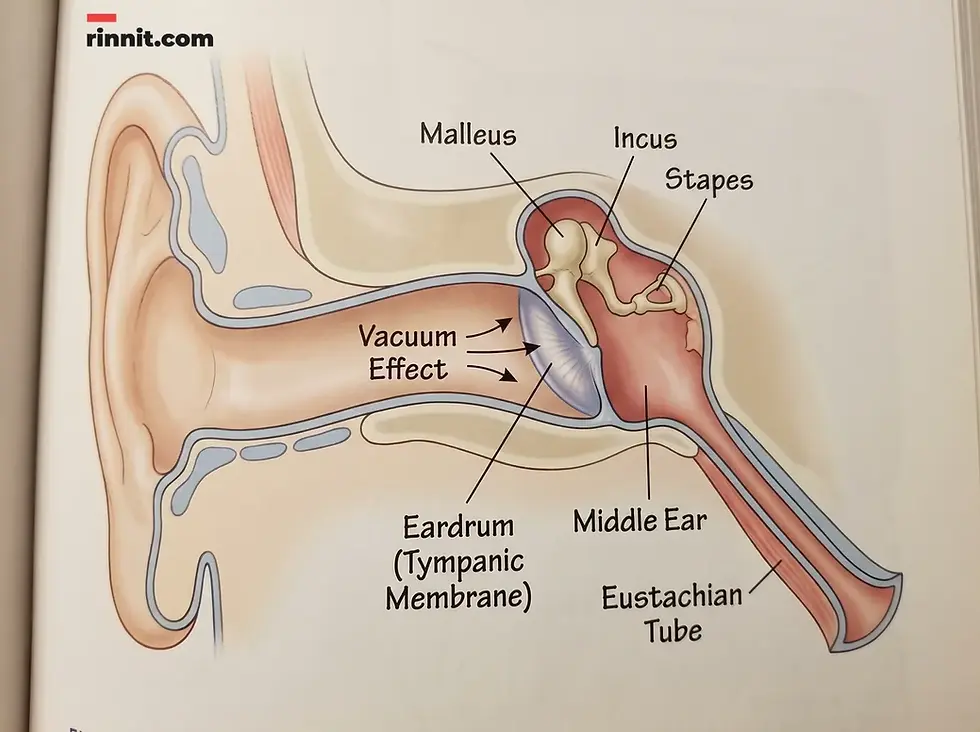
A retracted eardrum, clinically known as a retracted tympanic membrane, occurs when a disruption in air pressure balance sucks the eardrum toward the middle ear cavity. [1] Under normal conditions, the eardrum is cone-shaped with a slight natural dip in the center. [1] In a retracted state, this dip becomes significantly more pronounced as the membrane is pulled inward by a vacuum effect. [1]
The ear relies on equalized pressure between the external environment and the middle ear space. This balance is typically maintained by the eustachian tubes. When these tubes fail to function correctly, the resulting negative pressure pulls the eardrum out of its healthy alignment. [1] [3]
Feature | Normal Eardrum | Retracted Eardrum |
Shape | Cone-shaped with slight center dip | Deeply recessed toward middle ear |
Position | Suspended in middle ear space | May touch or stick to middle ear bones |
Function | Vibrates freely to transmit sound | Vibration may be limited, causing hearing loss |
Cause | Balanced internal/external pressure | Negative pressure vacuum in middle ear |
What are the symptoms and warning signs of a retracted eardrum?
Symptoms of a retracted eardrum often depend on the severity of the pull and whether the membrane has begun to press against the ossicles [1], the three tiny sound-conducting bones in the middle ear. In mild cases, an individual may not notice any symptoms at all [1], but as the condition progresses, several physical indicators often emerge.
Common signs include a persistent earache and a distinct feeling of fullness or pressure [1], similar to the sensation experienced during rapid altitude changes. If the eardrum's movement is restricted, temporary hearing loss may occur. [1] In some instances, fluid drainage from the ear, known as otorrhea, is present. [1] While permanent hearing loss is less common, it remains a risk if the retraction is severe and left untreated. [1]
What causes a retracted eardrum and who is at risk?
A retracted eardrum is mainly caused by eustachian tube dysfunction, when the tubes that equalize middle ear pressure become blocked or fail to open. [1] The eustachian tubes connect the middle ear to the upper throat [1], and their job is to open periodically to equalize pressure. If these tubes become blocked or fail to open, a vacuum forms, pulling the eardrum inward. Several underlying health conditions and environmental factors can contribute to this dysfunction:
Infections: Ear infections (otitis media), sinus infections, and upper respiratory infections [1] often cause inflammation that blocks the tubes.
Chronic Allergies: Long-term allergic reactions can lead to persistent swelling in the ear and throat tissues.
Structural Issues: Enlarged tonsils or adenoids [1] can physically obstruct the eustachian tube openings.
Pressure Changes: Rapid changes in altitude, such as during flying or diving, can trigger acute barotrauma and retraction.
What are the stages of eardrum retraction?
Healthcare providers use a specific staging system to categorize the severity of eardrum retraction. [1] This helps determine the appropriate level of monitoring or intervention required.
Stage | Description | Clinical Impact |
Stage 1 | Mild retraction | Eardrum is pulled back but does not touch the ossicles. |
Stage 2 | Moderate retraction | The membrane pulls far enough to touch the middle ear bones. |
Stage 3 | Severe retraction | The middle ear cavity begins to narrow significantly. |
Stage 4 | Adhesive retraction | Chronic inflammation causes the eardrum to become 'stuck' in the middle ear. |
What are the complications and long-term risks of a retracted eardrum?
If a retracted eardrum is not managed correctly, it can lead to more serious secondary conditions. One of the most significant risks is the development of a cholesteatoma, which is a noncancerous, cyst-like growth that forms behind the eardrum. [1] Over time, a cholesteatoma can expand and cause physical damage to the malleus, incus, and stapes bones. [1]
This damage to the ossicles interferes with the transmission of sound waves to the inner ear, potentially leading to permanent hearing loss. [1] Furthermore, the chronic inflammation associated with advanced stages can lead to permanent changes in the middle ear structure, making the condition harder to treat surgically.

How is a retracted eardrum diagnosed and managed?
Diagnosis is typically straightforward and involves a physical examination by an otolaryngologist (ENT). [1] Using an otoscope, a handheld tool with light and magnification, the provider can visually confirm the eardrum's position [1] and check for signs of inflammation or bone contact.
Monitoring and Self-Care
For mild (Stage 1) cases, providers often recommend a 'wait and see' approach. [1] The eardrum may return to its normal position as the underlying pressure issue resolves. [1] This process can take several weeks or even months. [1] During this period, the Valsalva maneuver, gently blowing air while pinching the nose and closing the mouth, may be recommended to help normalize ear pressure. [1]
Medical and Surgical Treatment
If the condition is severe or persistent, medical intervention becomes necessary to stabilize air pressure and protect hearing. [1]
Treatment Type | Method | Purpose |
Medication | Decongestants, Steroids, Antibiotics | Reduces inflammation, clears congestion, or treats infection. |
Ear Tubes | Tympanostomy | Small tubes are placed in the eardrum to provide constant pressure equalization. |
Tympanoplasty | Surgical repair | Repairs a ruptured or severely damaged eardrum membrane. |
Tonsillectomy or Adenoidectomy | Surgical removal | Removes enlarged tonsils or adenoids if they are blocking the eustachian tubes. |

Conclusion and Next Steps
A retracted eardrum is a common result of pressure imbalances within the ear, often linked to eustachian tube dysfunction. While many mild cases resolve with time and simple self-care, it is critical to monitor the condition closely. Timely diagnosis and intervention are essential to prevent complications such as cholesteatoma and permanent hearing loss. If you experience persistent ear fullness, pain, or muffled hearing, consult a healthcare professional to ensure your ear health is protected.
Next step: If you are experiencing persistent ear pressure or hearing changes, schedule an evaluation with an ENT specialist to prevent long-term complications.
Frequently Asked Questions
Can a retracted eardrum heal on its own?
Yes, mild cases often resolve without treatment as the underlying pressure imbalance corrects itself, though this may take several months.
What is the most common cause of eardrum retraction?
Eustachian tube dysfunction (ETD) is the most frequent cause, often triggered by infections, allergies, or structural blockages.
Is surgery always necessary for a retracted eardrum?
No. Surgery is typically reserved for severe cases where the eardrum touches the middle ear bones or interferes with hearing.
What are the risks of leaving a retracted eardrum untreated?
Untreated severe retraction can lead to cholesteatoma (a cyst-like growth), damage to the middle ear bones, and permanent hearing loss.
How do doctors stage eardrum retraction?
Doctors use a four-stage system based on how far the eardrum has pulled back and whether it is touching the middle ear bones or stuck in place.
Can flying make a retracted eardrum worse?
Yes, rapid changes in air pressure during flight can increase the risk of barotrauma and further retraction. Consult a doctor before flying if you have ear issues.
What does a retracted eardrum feel like?
Common sensations include a feeling of ear fullness, pressure, or a persistent earache, sometimes accompanied by muffled hearing.
What is the Valsalva maneuver?
The Valsalva maneuver is a self-care technique involving gently blowing air through the nose while it is pinched shut to help equalize pressure in the ears.
How long does it take for a retracted eardrum to improve?
Improvement can take anywhere from a few weeks to several months. If no progress is made within six months, treatment is usually recommended.
Can children get retracted eardrums?
Yes, children are particularly susceptible due to their smaller eustachian tubes and higher frequency of enlarged tonsils or adenoids.
Related Reading
Eustachian Tube Dysfunction: Symptoms, Types, Causes, Diagnosis, Treatment and Outlook — What You Need to Know explains the ear tube problem behind most retracted eardrums.
Cholesteatoma: What This Ear Growth Is, How It's Found, and How It's Removed explains the skin growth that can form behind a retracted eardrum.
Ear Tubes (Tympanostomy): Why They're Placed, What Happens, and How Recovery Goes explains how ear tubes are placed to let air into the middle ear.
Tympanoplasty: Eardrum Repair Surgery, Recovery, and Results explains surgery to repair or rebuild a damaged eardrum.
References
Cleveland Clinic. Retracted Eardrum. Cleveland Clinic. May 23, 2023. https://my.clevelandclinic.org/health/diseases/25019-retracted-eardrum
Cutajar J, Nowghani M, Tulsidas-Mahtani B, et al. The natural history of asymptomatic deep pars tensa retraction. The Journal of International Advanced Otology. 2018;14(1):10-14. PMID 29764774. https://doi.org/10.5152/iao.2018.5234
Urik M, Tedla M, Hurnik P. Pathogenesis of retraction pocket of the tympanic membrane: a narrative review. Medicina. 2021;57(5):425. PMID 33924803. https://doi.org/10.3390/medicina57050425
Spinos D, Mallick S, Judd O. Management of retraction pockets: historic and novel approaches. The Journal of Laryngology & Otology. 2022;136(7):582-587. PMID 34991748. https://doi.org/10.1017/S0022215121004321
Medical disclaimer
The information provided in this article is for educational purposes only and should not be used as a substitute for professional medical advice, diagnosis, or treatment. Always seek the advice of your physician or other qualified health provider with any questions you may have regarding a medical condition.



Comments