Renal Vein: Anatomy, Function, Conditions and Tests

Updated: 3 days ago
Medically reviewed by Dr. Baraa Alnahhal, MD · Last reviewed: October 2026
Quick Answer: What is the renal vein?
The renal veins are blood vessels that carry filtered, deoxygenated blood from each kidney to the inferior vena cava and on to the heart; the left renal vein is longer than the right. Conditions such as Nutcracker syndrome or renal vein thrombosis can block flow, causing severe pain or blood in the urine, and imaging evaluates them. [1]
What the research says
Left renal vein compression is common on imaging, but experts agree that imaging is needed to confirm Nutcracker syndrome. A modified Delphi consensus of 20 international venous-disease experts reached initial agreement on 24 of 37 statements. The report notes that asymptomatic compression is a common radiological finding; experts agreed that imaging is required to confirm Nutcracker syndrome but did not agree on specific diagnostic cut-off values (Heilijgers et al., Journal of Vascular Surgery: Venous and Lymphatic Disorders, 2025). [2]
Nutcracker syndrome is managed differently in children and adults. A clinical review describes compression of the left renal vein between the superior mesenteric artery and the aorta, which can cause hematuria, proteinuria, orthostatic hypotension, pain or even renal dysfunction. Conservative management is appropriate, especially in children, who tend to outgrow the disorder, while invasive options such as renal vein transposition or endovascular stenting are pursued more often in adults (Kolber et al., Cardiovascular Diagnosis and Therapy, 2021). [3]

What is the renal vein?
A renal vein is a blood vessel that carries blood from a kidney toward the heart. “Renal” means kidney. [1]
The kidneys filter waste products and extra fluid from the blood. Once filtration has occurred, renal veins carry the filtered, deoxygenated blood away from the kidneys and into the inferior vena cava. [1]
Most people have two kidneys, and each kidney has its own renal vein. The veins are not symmetrical: the right renal vein is shorter than the left. [1]

What do the renal veins do?
Renal veins are part of the circulatory system. Blood carries oxygen, hormones and nutrients to organs, muscles and tissues. It also helps remove waste products, including carbon dioxide, acids and excess water. [1]
Renal arteries bring oxygen-rich blood from the heart to the kidneys. The kidneys use oxygen and glucose as energy sources while filtering the blood. After filtration, the renal veins return deoxygenated, filtered blood to the heart. [1]
The renal veins empty into the inferior vena cava. This large vein carries deoxygenated blood from the lower part of the body toward the heart. [1]
Blood-flow step | What happens |
1. Renal arteries | Bring oxygenated blood from the heart to the kidneys |
2. Kidney filtration | The kidneys filter waste products and extra fluid from blood |
3. Renal veins | Carry filtered, deoxygenated blood away from the kidneys |
4. Inferior vena cava | Receives renal-vein blood and carries it toward the heart |
The renal veins support kidney circulation, but they do not perform filtration themselves. Their main role is to drain blood away from the kidneys after filtration. [1]
Where are the renal veins located?
The kidneys sit in the back of the abdomen, below the ribcage and above the waist. The renal veins lie near the kidneys and connect with the inferior vena cava. [1]
Left renal vein
The left renal vein lies behind the splenic vein and pancreas. It is farther from the inferior vena cava than the right renal vein, so it travels a longer distance. [1]
The left renal vein measures about 2 to 4 inches, or 6 to 10 centimeters, in length. [1]
Right renal vein
The right renal vein lies behind the top of the small intestine, also called the duodenum. It is closer to the inferior vena cava and is shorter. [1]
The right renal vein measures about three-quarters of an inch to 1.5 inches, or 2 to 4 centimeters, in length. [1]
Feature | Left renal vein | Right renal vein |
Side of body | Left | Right |
Relative length | Longer | Shorter |
Approximate length | 2 to 4 inches, or 6 to 10 centimeters | Three-quarters of an inch to 1.5 inches, or 2 to 4 centimeters |
Nearby structures | Behind the splenic vein and pancreas | Behind the top of the duodenum |
Typical drainage pattern | Kidney plus several nearby veins | Usually the right kidney alone |
Anatomic differences are normal. Length measurements are descriptive and are not diagnostic thresholds.
What drains into the left renal vein?
Because the left renal vein is longer and farther from the inferior vena cava, it receives blood from several nearby structures. [1]
Vein entering the left renal vein | Area it drains |
Left ureteral vein | The left ureter, which carries urine from the kidney to the bladder |
Left gonadal vein | The left ovary or testicle |
Left adrenal vein | The left adrenal gland, which produces hormones such as cortisol, epinephrine and norepinephrine |
Left inferior phrenic vein | The diaphragm, a muscle that helps with breathing |
The right renal vein usually receives blood only from the right kidney. Individual anatomy can vary, so an imaging report should be interpreted by a healthcare professional. [1]
Where does blood go after leaving the renal vein?
After the kidneys filter the blood, the renal veins send filtered, deoxygenated blood into the inferior vena cava. [1]
The inferior vena cava is a large vein that carries deoxygenated blood from the lower body to the heart. This route allows blood that has passed through the kidneys to rejoin the body’s broader circulation. [1]
Kidney filtration and venous drainage are connected but separate steps. The renal arteries bring blood into the kidneys, the kidneys filter it, and the renal veins carry the filtered blood away.
What conditions can affect renal veins?
Two conditions described in connection with renal veins are Nutcracker syndrome and renal vein thrombosis. [1]
Nutcracker syndrome
Nutcracker syndrome affects the left renal vein. The abdominal aorta and the superior mesenteric artery squeeze the left renal vein. This raises the blood pressure inside the vein and can compromise blood flow to the kidney. [1]
The name refers to the way the vein can be compressed between nearby structures. An imaging finding or symptom pattern does not establish this condition by itself.
Renal vein thrombosis
Renal vein thrombosis occurs when a blood clot forms in a renal vein. Conditions that may contribute include blood-clotting disorders, cancer and nephrotic syndrome.
A clot can affect blood flow through the renal vein. The significance depends on the person’s symptoms, the extent and location of the clot and the rest of the clinical picture.
Condition | What it involves |
Nutcracker syndrome | Compression of the left renal vein by the abdominal aorta and superior mesenteric artery |
Renal vein thrombosis | A blood clot forming in a renal vein |
Blood-clotting disorders | Conditions that may increase the tendency for a renal-vein clot |
Cancer | A condition that may contribute to renal vein thrombosis |
Nephrotic syndrome | A kidney condition that may contribute to renal vein thrombosis |
What happens if a renal vein is blocked?
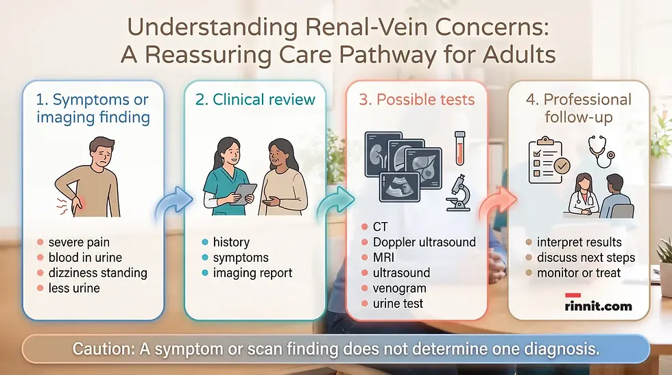
Renal-vein blockage may affect blood flow away from the kidney. Possible effects include severe pain, blood in the urine, dizziness when standing, male or female infertility, kidney damage or less urine than normal. [1]
A piece of a blood clot can also break free and travel to the lungs, causing a pulmonary embolism. This is a serious complication that requires immediate professional assessment. [1]
These symptoms have more than one possible cause. They should not be used to identify renal vein thrombosis or another condition without clinical evaluation.
What symptoms should be discussed with a healthcare professional?
Possible symptoms associated with renal-vein blockage include severe pain, blood in the urine, dizziness when standing, infertility concerns, kidney damage or reduced urine output. [1]
A person may not be able to tell whether a symptom involves a renal vein, the kidney itself or another structure. Promptly discuss severe or persistent symptoms with a healthcare professional, especially when urine output changes or blood appears in the urine.
If a clinician is evaluating a possible clot, share information about symptoms, timing, prior clotting problems, cancer, nephrotic syndrome, medicines and recent procedures when relevant.
How are renal veins evaluated?
The evaluation depends on the symptoms, medical history and question that needs to be answered. Imaging can show the renal veins and surrounding blood vessels, while urine testing can provide information about blood or protein in the urine.
Test or procedure | What it may help evaluate |
CT scan | Kidney and abdominal blood-vessel anatomy, including possible blockage or compression |
Duplex or Doppler ultrasound | Blood flow through vessels and possible changes in flow |
MRI | Detailed images of blood vessels and nearby structures |
Ultrasound | Kidney and abdominal anatomy without using X-rays |
Venogram | The inside of veins and venous blood flow using contrast imaging |
Urinalysis | Blood or high protein levels in urine |
High protein levels or blood in the urine may indicate kidney damage. A test list is not a personal testing plan; a healthcare professional can explain why a particular examination was chosen and what it can and cannot show. [1]
How can someone support general renal-vein health?
General care suggestions include having regular physical examinations to review blood-clot risk, taking prescribed blood-pressure medicines when recommended and using prescribed anticoagulant medicines when appropriate. [1]
Regular exercise can help keep blood moving. Other general suggestions include drinking adequate fluids, eating fruits and vegetables and quitting smoking. [1]
These are broad health-support measures, not a treatment plan for a clot or a blocked vein. Do not start, stop or change anticoagulants, blood-pressure medicines, fluids or exercise based only on an anatomy article.
General care focus | Practical conversation point |
Physical examinations | Ask how often your clot risk and kidney health should be reviewed |
Blood-pressure medicines | Take them only as prescribed and review concerns with a clinician |
Anticoagulants | Use blood thinners only when prescribed and monitored |
Movement | Choose regular activity that fits your abilities and health status |
Fluids | Ask what level of fluid intake is appropriate for you |
Fruits and vegetables | Discuss a suitable eating pattern if you have kidney or other medical conditions |
Tobacco | Seek professional support to quit smoking |
What questions should you ask a healthcare professional about the renal vein?
Useful questions about a renal-vein finding cover what it means in the imaging report, which test clarifies blood flow and which symptoms need immediate evaluation.
What does my renal-vein finding mean in the context of the full imaging report?
Is the concern compression, a blood clot, another vascular change or something else?
Does the finding involve the left renal vein, the right renal vein or both?
Which test is most useful for clarifying blood flow?
What do blood or protein in my urine mean in this context?
Could my symptoms have another cause outside the renal veins?
If an anticoagulant is being considered, what is its purpose and how will it be monitored?
What should I know about the risks of untreated blockage or a clot?
Which symptoms should prompt immediate evaluation?
What general changes to activity, fluids, smoking or diet are appropriate for me?
Conclusion: The kidneys’ return route to the heart
Renal veins are essential blood vessels that return filtered, deoxygenated blood from the kidneys to the heart. The left renal vein is longer and receives blood from several nearby veins, while the right renal vein is shorter and usually drains only the right kidney. [1]
Both renal veins empty into the inferior vena cava after kidney filtration. Nutcracker syndrome can compress the left renal vein, and renal vein thrombosis can form a clot that affects blood flow. [1]
Severe pain, blood in the urine, dizziness on standing, reduced urine or other concerns about kidney circulation deserve professional evaluation. CT, Doppler ultrasound, MRI, ultrasound, venogram and urine testing may help clinicians understand what is happening and plan appropriate follow-up. [1]
Frequently Asked Questions
What is the renal vein?
The renal vein is a blood vessel that carries filtered, deoxygenated blood from a kidney to the inferior vena cava and then toward the heart. Most people have one renal vein for each kidney.
Which renal vein is longer?
The left renal vein is longer. It is about 2 to 4 inches, or 6 to 10 centimeters, long. The right renal vein is about three-quarters of an inch to 1.5 inches, or 2 to 4 centimeters, long.
Why is the left renal vein longer?
The left renal vein is farther from the inferior vena cava, so it travels a longer distance. It lies behind the splenic vein and pancreas.
What drains into the left renal vein?
The left renal vein may receive blood from the left ureteral, gonadal, adrenal and inferior phrenic veins in addition to blood from the left kidney.
What is Nutcracker syndrome?
Nutcracker syndrome involves the abdominal aorta and superior mesenteric artery squeezing the left renal vein. The compression raises pressure inside the vein and can compromise blood flow to the kidney.
What is renal vein thrombosis?
Renal vein thrombosis is a blood clot in a renal vein. Blood-clotting disorders, cancer and nephrotic syndrome may contribute to clot formation.
What are possible signs of renal-vein blockage?
Possible effects include severe pain, blood in the urine, dizziness when standing, infertility, kidney damage, reduced urine or a clot fragment traveling to the lungs as a pulmonary embolism. These effects are not present in every case and do not establish a diagnosis by themselves.
What tests can examine the renal veins?
Possible tests include CT, duplex or Doppler ultrasound, MRI, ultrasound and venogram. A urine test may look for blood or high protein levels that can indicate kidney damage.
Are blood thinners always needed for renal-vein problems?
No. Anticoagulants are prescribed only in selected situations. The need for a blood thinner depends on the specific condition and should be decided and monitored by a healthcare professional.
What symptoms can Nutcracker syndrome cause?
Possible symptoms include blood in the urine, belly or flank pain and dizziness when standing. Many people whose left renal vein looks squeezed on imaging have no symptoms, so a specialist confirms the diagnosis.
Related Reading
Nutcracker Syndrome: Understanding Left Renal Vein Entrapment explains how the left renal vein gets squeezed and how it is treated.
Understanding Renal Vein Thrombosis: Symptoms, Causes, and Care covers blood clots in the kidney veins and their treatment.
Nephrotic Syndrome: Symptoms, Causes, and Treatment Guide explains a kidney condition that raises the risk of renal vein clots.
Pulmonary Embolism Guide: Symptoms, Causes, Diagnosis, and Treatment describes the emergency when a blood clot travels to the lungs.
References
Cleveland Clinic. Renal Vein: Anatomy & Function. Cleveland Clinic. Last updated October 3, 2024. https://my.clevelandclinic.org/health/body/renal-vein
Heilijgers F, Gloviczki P, O’Sullivan G, et al. Nutcracker syndrome (a Delphi consensus). J Vasc Surg Venous Lymphat Disord. 2025;13(1):101970. PMID 39362632. https://doi.org/10.1016/j.jvsv.2024.101970
Kolber MK, Cui Z, Chen CK, et al. Nutcracker syndrome: diagnosis and therapy. Cardiovasc Diagn Ther. 2021;11(5):1140-1149. PMID 34815965. https://doi.org/10.21037/cdt-20-160
Disclaimer
This article is for general educational purposes and is not a substitute for professional medical advice, diagnosis or treatment. Always seek the advice of a qualified health provider with any questions you may have regarding a medical condition.
It explains renal-vein anatomy, blood flow, conditions, symptoms and evaluation topics in clear language. It does not determine what an individual symptom or imaging finding means, and it does not replace evaluation by a qualified healthcare professional. Seek urgent help for sudden severe pain, chest pain, trouble breathing or other possible emergencies.



Comments