Renal Sinus: Anatomy, Function, Cysts and Imaging

Updated: 3 days ago
Medically reviewed by Dr. Baraa Alnahhal, MD · Last reviewed: October 2026
Quick Answer: What is the renal sinus?
The renal sinus is a fat-filled cavity in the middle of each kidney that cushions important structures and contains the renal artery and vein, renal pelvis, calyces, nerves and lymphatic channels. It is normal anatomy, not a diagnosis. Simple cysts there often cause no symptoms. Pain, fever or blood in the urine should be discussed with a professional. [1]
What the research says
Fat in the renal sinus has been linked to blood pressure control. In 205 adults aged 55 to 85 at risk for cardiovascular events, MRI-measured renal sinus fat correlated with the number of prescribed antihypertensive medications (P = 0.010) and with stage II hypertension (P = 0.02), after accounting for age, sex, height, body mass index and intraperitoneal fat. The study shows an association and cannot prove that renal sinus fat causes hypertension (Chughtai et al., Hypertension, 2010). [2]
Kidney fat measured on imaging is higher in people with metabolic conditions. A systematic review of 28 studies (6,994 participants) found higher renal fat fractions in people with obesity, diabetes, chronic kidney disease and hypertension. MRI was used in 75% of the studies, CT was limited to renal sinus evaluation, and the authors noted that imaging methods still need standardization (Raphael et al., Journal of Clinical Medicine, 2025). [3]

What is the renal sinus?
The renal sinus is a central, fat-filled space inside the kidney. Think of it as a protected compartment that holds several structures needed for blood flow, nerve signaling, lymphatic drainage and urine collection. [1]
Most of the renal sinus is fat. The fat surrounds and cushions the structures inside it. The space is not an abnormal growth or a disease by itself; it is part of normal kidney anatomy. [1]

What does the renal sinus do?
The renal sinus acts as a compartment and cushion. It contains the kidney’s vascular system, including arteries, veins and the tiny capillaries that connect them. It also contains part of the kidney’s nervous system and lymphatic channels. [1]
Blood enters the kidney through the renal artery. The kidney filters waste products and excess fluid from the blood. Those waste products and extra fluid eventually leave the body as urine. [1]
The renal pelvis and calyces support the urine pathway. The calyces are where urine collection begins, and the renal pelvis gathers urine before it moves into the ureter. [1]
Renal sinus structure | Role described in kidney anatomy |
Fat | Wraps around and cushions the other structures |
Renal pelvis | Collects urine as the kidneys produce it |
Major and minor calyces | Begin urine collection |
Renal artery | Brings blood into the kidney |
Renal vein | Carries blood away from the kidney |
Autonomic nerves | Help regulate involuntary actions |
Lymphatic channels | Provide lymphatic drainage pathways |
Connective tissue | Supports the structures within the compartment |
The amount of fat in the renal sinus gradually increases with age and weight gain. That observation does not diagnose a kidney problem in a particular person.
Where is the renal sinus located?
The renal sinus is in the middle of the kidney. Renal parenchyma lies along its sides. The parenchyma includes the renal cortex and renal medulla, two major regions involved in kidney function. [1]
The renal hilum sits at the kidney’s entry-and-exit area. Blood vessels, nerves, lymphatic channels and the ureter pass through the hilum to enter or leave the renal sinus. [1]
Renal sinus, renal pelvis and renal hilum
These terms describe related but different parts of the kidney.
Term | Simple explanation |
Renal sinus | The central, fat-filled compartment that contains several kidney structures |
Renal pelvis | A urine-collecting space inside the renal sinus |
Renal hilum | The entry-and-exit area where vessels, nerves, lymphatic channels and the ureter pass through |
Renal parenchyma | The kidney tissue around the renal sinus, including the cortex and medulla |
How does the renal sinus appear on an ultrasound?
The renal sinus is padded mostly with fat, which usually appears white or yellow in ordinary anatomy illustrations. [1]
On an ultrasound, the renal sinus often looks hyperechoic compared with the renal parenchyma. Hyperechoic means that the tissue reflects many ultrasound sound waves. It commonly appears light gray and may look round or oval. [1]
An ultrasound appearance is one part of an imaging examination. A description on an imaging report should be interpreted with the rest of the report and the person’s clinical history rather than used for self-diagnosis.
What conditions can affect the renal sinus?
Conditions such as caliectasis and vascular findings can involve the renal sinus or the structures within it. A finding in this area does not automatically mean cancer, and a symptom does not identify one particular condition. [1]
Condition or finding | Description |
Renal sinus cyst | A benign, fluid-filled sac that may grow inside or outside the renal sinus |
Parapelvic cyst | A cyst that grows outside the renal sinus |
Peripelvic cyst | A cyst that grows inside the renal sinus |
Renal sinus lipomatosis | An excess amount of fat in the renal sinus |
Benign multilocular cystic nephroma | A noncancerous tumor that can grow into the renal sinus |
Caliectasis | Swelling of the calyces with extra fluid |
Vascular lesions | Changes involving a blood-vessel wall or an unusual vascular connection |
Renal cell carcinoma | A cancerous tumor that can grow into the renal sinus |
Caliectasis affects the urine-collection system. Without treatment, it may cause kidney failure. Vascular findings may include a renal artery aneurysm, an arteriovenous communication or a renal vein varix. [1]
The amount of fat within the renal sinus may also affect blood pressure. This relationship is not a personal diagnosis or a reason to change medication without professional guidance.
What are renal sinus cysts?
A renal sinus cyst is a benign, fluid-filled sac. It may be located inside the renal sinus or just outside it. Simple renal cysts usually do not cause symptoms, and many people do not know they have one. [1]
Cysts are often found during an imaging exam performed for another health reason. A healthcare professional may recommend regular check-ups or repeat imaging to monitor a cyst. [1]
Cysts and lipomatosis are common types of renal sinus lesions with aging. About 1 in 3 people over age 70 has at least one renal cyst, which may be inside or outside the renal sinus. This is a population statistic, not an individual prediction.
Surgery is uncommon. It may be considered if a cyst bursts, bleeds, becomes too large or becomes infected. [1]
What is renal sinus invasion?
Renal sinus invasion means that a cancerous tumor has spread into the renal sinus. Renal cell carcinoma is the most common cancer associated with renal sinus invasion. [1]
A CT scan can usually diagnose renal sinus invasion. A finding described as possible invasion requires professional interpretation alongside the imaging details and the rest of the clinical evaluation. [1]
How is renal sinus lipomatosis evaluated?
A CT scan can identify fatty masses in the renal sinus. CT or MRI can usually confirm that the extra tissue is fat, which is characteristic of renal sinus lipomatosis.
Surgery or a biopsy is needed only when imaging suggests something other than ordinary fat. This does not mean that every increase in renal sinus fat requires surgery; the next step depends on the finding and the clinical context.
What symptoms may occur?
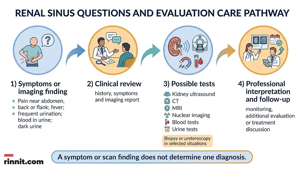
Many simple renal sinus cysts cause no symptoms. When a renal sinus condition or a related kidney problem is causing trouble, symptoms may include pain in the stomach, back or the area between the ribs and hip, fever, frequent urination, blood in the urine or dark urine.
These symptoms can have many possible causes. They do not identify a renal sinus cyst, lipomatosis, caliectasis, vascular lesion or tumor by themselves.
Contact a healthcare professional if you have kidney-related symptoms, especially if they persist, recur or occur together. Bring the imaging report, a list of medicines and a brief timeline of symptoms if those details are available.
How is the renal sinus examined?
Healthcare professionals choose tests based on the finding, symptoms, medical history and the question that needs to be answered.
Test or procedure | What it may help examine |
Kidney ultrasound | Kidney structure and the appearance of the renal sinus without using X-rays |
CT scan | Kidney abnormalities, growths, fatty masses or possible renal sinus invasion |
MRI | Detailed images of kidney structures and abnormalities |
Nuclear imaging | Kidney structure or function using a nuclear-medicine study |
Blood tests | Aspects of kidney health and overall clinical evaluation |
Urine tests | Blood, other urine findings and kidney-health clues |
Kidney biopsy | Tissue examined when a biopsy is considered appropriate |
Ureteroscopy | Direct examination of parts of the urinary tract in selected situations |
A test list is not a personal testing plan. A healthcare professional can explain why a particular examination was recommended and what its limitations are.
How can someone support general kidney health?
General kidney-health guidance includes maintaining a healthy weight for you, quitting smoking, reducing salt, drinking plenty of water and getting at least 30 minutes of physical activity five days a week. [1]
General guidance also advises limiting regular NSAID use, keeping an eye on blood pressure and monitoring blood sugar when diabetes is present. These are general health behaviors, not a personalized treatment plan. [1]
If you have kidney disease, high blood pressure, diabetes, take NSAIDs regularly or have been told to restrict fluids, ask a healthcare professional which habits are appropriate for you.
General health focus | Practical conversation point |
Healthy weight | Ask what a sustainable weight goal means for you |
Tobacco | Seek help to quit if you smoke |
Salt | Ask how much salt is appropriate for your health needs |
Fluids | Discuss an appropriate fluid pattern if you have kidney or heart conditions |
Physical activity | Aim for activity that fits your abilities and health status |
NSAIDs | Review regular use with a healthcare professional |
Blood pressure | Monitor it as recommended |
Blood sugar | Monitor it if diabetes is present |
Do not start, stop or change a medicine based only on an anatomy article or an imaging phrase.
What questions should you ask a healthcare professional about the renal sinus?
Useful questions about a renal sinus finding cover what it means in the full imaging report, whether follow-up imaging is needed and which symptoms need faster evaluation.
What does the renal sinus finding mean in the context of the full imaging report?
Is the finding a cyst, a fatty mass, a urine-collection change, a vascular finding or something else?
Is the renal pelvis involved, or is the finding limited to the surrounding renal sinus?
Do I need repeat imaging or a follow-up appointment?
Which test, if any, would clarify the finding?
Could my pain, fever, urinary frequency, blood in the urine or dark urine have another cause?
Should I change anything about NSAID use, salt, fluids or physical activity?
If a procedure is being considered, what is its purpose and what are its risks?
Which symptoms should prompt faster evaluation?
Conclusion: A normal compartment with a protective role
The renal sinus is a normal, fat-filled compartment in the center of the kidney. It cushions and contains the renal pelvis, calyces, blood vessels, nerves, lymphatic channels, connective tissue and surrounding fat. [1]
Its relationship with the renal pelvis and renal hilum can help make kidney anatomy and imaging language easier to understand. Renal sinus cysts are common findings, and simple cysts often cause no symptoms. Other findings, including lipomatosis, caliectasis, vascular lesions or a growth, need interpretation in context. [1]
Pain near the abdomen, back or flank, fever, frequent urination, blood in the urine or dark urine deserves professional attention. A healthcare professional can connect symptoms with the appropriate imaging, laboratory tests, follow-up or treatment discussion. [1]
Frequently Asked Questions
Is the renal sinus a disease?
No. The renal sinus is a normal central compartment in the kidney. It contains fat, the renal pelvis, calyces, blood vessels, nerves, lymphatic channels and connective tissue. Conditions can affect the renal sinus, but the anatomical term itself is not a diagnosis.
What is the difference between the renal sinus and the renal pelvis?
The renal pelvis is inside the renal sinus. It is a larger space that collects urine as the kidneys make it. Urine then flows from the renal pelvis into the ureter.
What is the renal hilum?
The renal hilum is the kidney’s entry-and-exit area. Blood vessels, nerves, lymphatic channels and the ureter pass through the hilum to enter or leave the renal sinus.
Do renal sinus cysts always need surgery?
No. Simple renal cysts usually do not cause symptoms and are often monitored with check-ups or imaging. Surgery is uncommon but may be needed if a cyst bursts, bleeds, becomes too large or becomes infected.
What tests can examine the renal sinus?
Possible examinations include kidney ultrasound, CT, MRI and nuclear imaging. Blood tests, urine tests, kidney biopsy and ureteroscopy may also be used in selected situations. The appropriate plan depends on the finding and the person’s symptoms and history.
Does a renal sinus finding mean cancer?
No. Renal sinus cysts are benign, and several other noncancerous findings can affect this area. Renal cell carcinoma can grow into the renal sinus, but an imaging finding requires professional interpretation and does not establish cancer by itself.
What symptoms should be discussed with a healthcare professional?
Discuss pain in the stomach, back or between the ribs and hip, fever, frequent urination, blood in the urine or dark urine. These symptoms can have different causes and do not identify one renal sinus condition.
What is inside the renal sinus?
The renal sinus is mostly fat. It also holds the renal pelvis, calyces, blood vessels, nerves, lymphatic channels and connective tissue.
What is renal sinus lipomatosis?
Renal sinus lipomatosis means there is extra fat in the renal sinus. Fat in this area tends to increase with age and weight gain, and a CT or MRI can usually confirm that the tissue is fat.
Why does the renal sinus look bright on an ultrasound?
The renal sinus is mostly fat, which reflects many sound waves. This makes it look lighter than the surrounding kidney tissue, which is a normal finding called hyperechoic.
Related Reading
Kidney Cysts: Symptoms, Causes, Diagnosis, and Treatment Options explains simple and complex kidney cysts and when they need care.
Hydronephrosis: What Causes Kidney Swelling, How It Is Found, and How It Is Treated explains kidney swelling caused by blocked urine flow.
Kidney Ultrasound: Why It's Done, How to Prepare and What It Shows describes the imaging test that often shows the renal sinus.
Kidney Cancer: Symptoms, Causes & Treatment Guide covers the warning signs, staging and treatment of kidney cancer.
References
Cleveland Clinic. Renal Sinus: Anatomy, Function & Complications. Cleveland Clinic. Last updated May 22, 2026. https://my.clevelandclinic.org/health/body/24182-renal-sinus
Chughtai HL, Morgan TM, Rocco M, et al. Renal sinus fat and poor blood pressure control in middle-aged and elderly individuals at risk for cardiovascular events. Hypertension. 2010;56(5):901-906. PMID 20837881. https://doi.org/10.1161/HYPERTENSIONAHA.110.157370
Raphael H, Klang E, Konen E, et al. Renal fat accumulation assessed by MRI or CT and its association with clinical and metabolic disorders: a systematic imaging review. J Clin Med. 2025;14(12):4305. PMID 40566049. https://doi.org/10.3390/jcm14124305
Disclaimer
This article is for general educational purposes and is not a substitute for professional medical advice, diagnosis or treatment. Always seek the advice of a qualified health provider with any questions you may have regarding a medical condition.
It explains renal sinus anatomy, common findings, symptoms and evaluation topics in clear language. It does not determine what an individual imaging finding means or replace an evaluation by a qualified healthcare professional.



Comments