Radius Bone: Anatomy, Location, Function and Common Problems

Updated: 3 days ago
Medically reviewed by Dr. Baraa Alnahhal, MD · Last reviewed: October 2026
Quick Answer: What is the radius bone?
The radius is one of the two forearm bones, lying on the thumb side opposite the ulna, and helps the arm and wrist move, flex and rotate. Its upper end meets the humerus at the elbow, and its lower end helps form the wrist joint. Fractures and osteoporosis can affect it; seek emergency care for a suspected fracture. [1]
What the research says
For older adults with a wrist fracture, surgery brings small functional gains and a different complication profile than a cast. A network meta-analysis of 23 randomized trials (2,020 patients older than 60 with distal radius fractures) found that volar locking plate fixation improved patient-rated wrist scores (mean difference −4.45 points) and grip strength (6.11%) compared with cast immobilization, but most differences were not clinically relevant. Plating had the lowest rate of minor complications and one of the highest rates of major complications. Treatment choice is individual (Gutiérrez-Espinoza et al., International Journal of Environmental Research and Public Health, 2023). [4]
Nursemaid elbow in young children is treated with a quick clinician-performed maneuver, and one maneuver works better. A systematic review and meta-analysis of 7 randomized trials (701 children) found hyperpronation was more effective than supination–flexion (risk ratio 0.34, 95% CI 0.23–0.49; absolute difference 26%; number needed to treat 4). This is a procedure for healthcare professionals, not something to try at home (Bexkens et al., The American Journal of Emergency Medicine, 2017). [5]

What is the radius bone?
The radius is a long bone in the forearm. Every forearm has a radius and an ulna. Together, these bones support movement of the arm, wrist and hand. [1]
The radius also provides attachment and support for important muscles, tendons, ligaments and blood vessels. Because so many structures connect with it, an injury to the radius can affect nearby muscles and nerves as well. [1]
If the radius breaks, treatment may involve a splint or cast. Some fractures need surgery to realign the bone, followed by physical therapy to help restore strength and movement.
What does the radius do?
The radius has several important jobs. It helps the forearm and wrist move, flex and rotate. It also holds seven muscles in place and helps stabilize the rest of the arm, wrist and hand. [1]
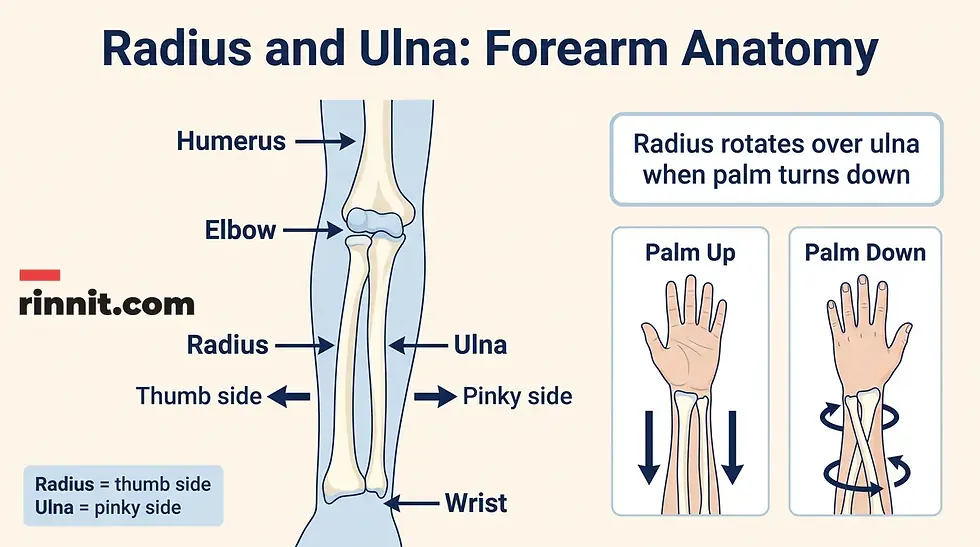
The radius and ulna work together to allow the forearm to turn. When the arm is extended with the palm facing down, the radius rotates over the ulna. When the palm faces up, the two bones are more parallel. [1]
Function | Why it matters |
Movement | Helps the forearm and wrist move, flex and rotate. |
Muscle support | Holds seven muscles in place. |
Stability | Helps stabilize the arm, wrist and hand. |
Rotation | Moves over the ulna as the forearm turns palm-down. |
Where is the radius located?
The radius is on the lateral, or thumb-side, of the forearm. The ulna lies on the medial, or pinky-side, of the forearm. [1]
The radius begins at the elbow, where its upper end meets the humerus. It continues down the forearm toward the wrist, where its wider lower end meets two wrist bones called the scaphoid and lunate. [1]
The radius and ulna are not simply parallel rods. Their shapes and connections allow the wrist and hand to rotate smoothly. Their relationship is why the palm can turn from facing up to facing down.
What does the radius look like?
The radius has a small upper end, a long middle shaft and a wider lower end. It is slightly curved, thicker and slightly shorter than the ulna. [1]
Proximal radius
The proximal, or upper, end of the radius connects with the humerus at the elbow. It includes the head, neck and radial tuberosity. [1]
Radius shaft
The shaft is the long middle section. It supports the weight of the forearm and helps give the forearm its shape.
Distal radius
The distal, or lower, end is wider than the shaft. It forms the top of the wrist joint and meets the scaphoid and lunate wrist bones. It includes the styloid process and ulnar notch. [1]
Part | Plain-language description |
Head | Part of the upper radius near the elbow. |
Neck | Narrow section below the radial head. |
Radial tuberosity | Structure on the upper radius. |
Shaft | Long middle section of the bone. |
Styloid process | Feature of the lower radius near the wrist. |
Ulnar notch | Area of the distal radius that relates to the ulna. |
These terms help healthcare providers describe where pain, injury or a fracture is located. They are not labels you need to use to diagnose yourself.
How big is the radius?
The radius is one of the longest bones in the body. Most adults’ radius bones are around 10 inches (25 centimeters) long. [1]
Individual bone size varies. A measurement alone does not indicate whether a bone is healthy or injured.
What problems can affect the radius?
The most common problems described for the radius are fractures, osteoporosis and damage to nearby muscles or nerves.
Problem | What may happen |
Radius fracture | The bone breaks, often after a fall, car accident or sports injury. |
Osteoporosis | Bone becomes weaker and more vulnerable to sudden, unexpected fractures. |
Nearby muscle or nerve injury | Structures attached to or connected with the radius may also be affected. |
Nursemaid elbow | In a young child, the radius can partially dislocate where it meets the elbow. |
A symptom does not identify the exact problem by itself. A healthcare provider can assess the injury, examine movement and choose appropriate testing when needed.
What are the symptoms and causes of radius fractures?
A radius fracture is a broken radius. Common causes include falls, car accidents and sports injuries.
Possible symptoms include pain, swelling, tenderness, bruising or discoloration, and an inability to move the arm as usual. A deformity or bump that was not there before the injury can also occur.
Some named fracture patterns include Colles fractures, Smith fractures, greenstick fractures and growth-plate fractures.

When is a radius fracture an emergency?
Go to an emergency room right away if you have experienced trauma or think you may have a fracture. Do not wait for symptoms to become more severe before seeking evaluation.
How does osteoporosis affect the radius bone?
Osteoporosis weakens bones and makes them more susceptible to sudden and unexpected fractures. It often has no noticeable symptoms, so some people do not know they have it until a bone breaks. Fractures most often happen in the hip, the spine and the wrist, according to the National Institute of Arthritis and Musculoskeletal and Skin Diseases. [3]
Females and adults older than 65 have an increased risk of developing osteoporosis. If you are concerned about bone health, a healthcare provider can discuss whether a bone-density test is appropriate. MedlinePlus offers a plain-language overview of risk factors, screening and treatment.
A bone-density test, sometimes called a DEXA or DXA scan, uses low levels of X-rays to measure bone strength and track bone loss as you age.
What is nursemaid elbow in children, and how does it involve the radius?
Nursemaid elbow, also called pulled elbow, is a common injury in young children. It is a partial dislocation of the radius where it meets the elbow.
Children’s ligaments are looser than adults’ ligaments. Pulling on a child’s arm or hand can cause this injury, sometimes during an accidental tug or when a child is lifted by the hands.
If a young child suddenly stops using an arm after a pull or tug, contact a healthcare provider for guidance rather than trying to put the elbow back into place yourself.
What tests may be used?
A bone-density test can help evaluate bone strength and bone loss over time. When a provider needs to assess a suspected fracture or other injury, imaging may include X-rays, MRI or a CT scan.
The appropriate test depends on the problem being evaluated. Imaging should be selected and interpreted by a healthcare professional.
Test | General role described |
Bone-density test / DEXA / DXA | Measures bone strength and tracks bone loss. |
X-ray | Provides imaging of bone and possible fracture or injury. |
MRI | Provides detailed imaging when further assessment is needed. |
CT scan | Provides cross-sectional imaging when further assessment is needed. |
How are radius problems treated?
Treatment depends on the condition and the injury pattern. A radius fracture may need immobilization with a splint or cast. Some fractures need surgery to realign the bone so it can heal. Physical therapy may help restore strength and movement afterward.
Osteoporosis treatment can include exercise, vitamin and mineral supplements and medications. A healthcare provider can develop a plan tailored to your bone health. [3]
Do not use this overview to choose a treatment or change medicines on your own. A fracture, suspected osteoporosis or a child’s elbow injury needs individualized professional care.
How can you support bone health?
General habits that support bone and overall health include following a nutritious diet and exercise plan and attending regular checkups.
Safety steps can reduce the risk of injury. Wear a seatbelt, use appropriate protective equipment during activities and sports, keep your home and workspace free of clutter that could cause a trip, and use proper tools rather than standing on chairs, tables or countertops.
If walking is difficult or your fall risk is higher, use a cane or walker as advised. If you are older than 50 or have a family history of osteoporosis, ask a healthcare provider whether a bone-density scan may be appropriate.
Radius and ulna: what is the difference?
The radius and ulna are the two bones of the forearm. The radius is on the thumb side, while the ulna is on the pinky side. The ulna is slightly longer than the radius. [1]
They share the work of supporting forearm and wrist movement. Their shape and connection allow the hand and wrist to rotate smoothly as the palm turns up or down.
Neither bone is more important than the other. They work as a coordinated pair.
Conclusion: A thumb-side bone with a big job
The radius is a long, thumb-side forearm bone that helps the wrist and forearm move, flex and rotate. It supports muscles and other structures, meets the humerus at the elbow and forms part of the wrist joint at its wider lower end.
Falls, sports injuries and car accidents can fracture the radius. Osteoporosis can weaken it and increase the chance of an unexpected fracture. Pain, swelling, tenderness, bruising, reduced movement or a new deformity after trauma deserves prompt attention, and suspected fractures require emergency evaluation.
If you are concerned about radius pain, a forearm injury or bone strength, note when the problem started and which movements are affected, then contact a healthcare provider for guidance.
Frequently Asked Questions
Is the radius the same as the ulna?
No. The radius and ulna are the two different bones in the forearm. The radius is on the thumb side, and the ulna is on the pinky side.
Which bone is longer, the radius or ulna?
The ulna is slightly longer than the radius. The two bones work together to support forearm and wrist movement.
Does the radius rotate?
Yes. When the palm turns downward with the arm extended, the radius rotates over the ulna. The bones are more parallel when the palm faces upward.
Where does the radius meet the wrist?
The wider distal end of the radius forms the top of the wrist joint and meets the scaphoid and lunate wrist bones.
What are common signs of a radius fracture?
Pain, swelling, tenderness, bruising or discoloration, difficulty moving the arm as usual and an unusual bump or deformity can occur. If you have experienced trauma or think you have a fracture, go to an emergency room right away.
Can osteoporosis affect the radius?
Yes. Osteoporosis weakens bones and can make the radius more susceptible to sudden and unexpected fractures. A healthcare provider can discuss bone-density testing when appropriate.
What is nursemaid elbow?
Nursemaid elbow is a partial dislocation of the radius at a young child’s elbow. It can happen after pulling on a child’s arm or hand because children’s ligaments are looser than adults’ ligaments.
Can a radius fracture need surgery?
Some radius fractures need surgery to realign the bone, while others may be treated with immobilization such as a splint or cast. The appropriate plan depends on the fracture and should be determined by a healthcare professional.
What is a Colles fracture?
A Colles fracture is a break near the wrist end of the radius in which the broken piece tilts toward the back of the hand. It often happens after a fall onto an outstretched hand.
How long is the radius bone?
In most adults, the radius is about 10 inches (25 centimeters) long. Bone size varies from person to person.
Related Reading
Broken Wrist: Symptoms, Causes, and Treatment explains wrist fractures, many of which involve the radius.
What Is a Colles Fracture? Symptoms, Causes, Diagnosis and Treatment covers the common break at the wrist end of the radius.
The Ulna Bone: Anatomy, Function, and Common Conditions describes the forearm bone that works alongside the radius.
Osteoporosis: Causes, Symptoms, Diagnosis and Treatment Explained explains bone thinning that makes wrist fractures more likely.
References
Cleveland Clinic. Radius Bone: What It Is, Anatomy, Location & Function. Cleveland Clinic. Last updated August 4, 2026. https://my.clevelandclinic.org/health/body/24528-radius
National Library of Medicine. Osteoporosis. MedlinePlus. Last updated September 30, 2025. https://medlineplus.gov/osteoporosis.html
National Institute of Arthritis and Musculoskeletal and Skin Diseases. Osteoporosis. NIAMS. Last reviewed December 2022. https://www.niams.nih.gov/health-topics/osteoporosis
Gutiérrez-Espinoza H, Araya-Quintanilla F, Cuyul-Vásquez I, et al. Effectiveness and safety of different treatment modalities for patients older than 60 years with distal radius fracture: a network meta-analysis of clinical trials. Int J Environ Res Public Health. 2023;20(4):3697. PMID 36834390. https://doi.org/10.3390/ijerph20043697
Bexkens R, Washburn FJ, Eygendaal D, et al. Effectiveness of reduction maneuvers in the treatment of nursemaid’s elbow: a systematic review and meta-analysis. Am J Emerg Med. 2017;35(1):159-163. PMID 27836316. https://doi.org/10.1016/j.ajem.2016.10.059
Disclaimer
This article is for general educational purposes and is not a substitute for professional medical advice, diagnosis or treatment. Always seek the advice of a qualified health provider with any questions you may have regarding a medical condition.
It explains radius anatomy, function, common problems, testing and general care considerations in plain language. It is non-diagnostic and does not determine whether an individual has a fracture or osteoporosis or replace an examination by a qualified healthcare professional.



Comments