Pleura: Anatomy, Function, Conditions and Care

Updated: 3 days ago
Medically reviewed by Dr. Baraa Alnahhal, MD · Last reviewed: October 2026
Quick Answer: What is the pleura?
The pleura is a two-layered membrane around the lungs and along the chest wall. Its fluid-filled pleural space lets the layers glide during breathing, while the membrane cushions the lungs and helps prevent friction against the chest wall. Its two layers are the visceral pleura and the parietal pleura. [1]
Core idea | Plain-language meaning |
What it is | A thin, watery serous membrane. |
Where it is | Along the chest wall and around the lungs. |
Layers | Visceral pleura and parietal pleura. |
Between the layers | A pleural space containing a thin layer of pleural fluid. |
Main role | Cushioning the lungs and reducing rubbing during breathing. |
Concerning symptoms | Sharp breathing-related pain or dyspnea. |
What the research says
Ultrasound guidance may make thoracentesis safer. A systematic review and meta-analysis of three randomized trials (417 patients) found that ultrasound-guided thoracentesis was successful in 195 of 202 patients (96.5%), compared with 189 of 215 (87.9%) using the anatomic landmark approach. In the two studies that assessed it, pneumothorax occurred in 1 of 99 patients in the ultrasound group and 20 of 113 in the landmark group. The differences in success (OR 3.99, 95% CI 0.60–26.50) and complications (OR 0.18, 95% CI 0.01–3.07) only trended toward benefit, with wide confidence intervals (Lakkadghatwala et al., Am J Emerg Med, 2025). [2]
Some first-episode pneumothoraces can be observed rather than drained. In a multicenter randomized noninferiority trial of 316 patients aged 14 to 50 with a first moderate-to-large primary spontaneous pneumothorax, lung re-expansion within 8 weeks occurred in 98.5% (129 of 131) with interventional management and 94.4% (118 of 125) with conservative management (risk difference −4.1 percentage points, 95% CI −8.6 to 0.5). In the conservative group, 15.4% (25 of 162) eventually needed an intervention, and conservative management carried a lower risk of serious adverse events or recurrence. The authors noted that the result was not statistically robust to conservative assumptions about missing data (Brown et al., N Engl J Med, 2020). [3]
For malignant pleural effusion, a catheter and pleurodesis trade different benefits and risks. A systematic review and meta-analysis of five randomized trials (545 patients) found no differences in survival or breathlessness between an indwelling pleural catheter and chemical pleurodesis. The catheter was associated with a shorter hospital stay and fewer repeat pleural procedures (RR 0.32, 95% CI 0.18–0.55) but a higher risk of cellulitis (RR 5.83, 95% CI 1.56–21.8) (Iyer et al., Ann Am Thorac Soc, 2019). [4]

What is the pleura?
The pleura is a thin, watery tissue called a serous membrane. It lines the walls of the chest, or thorax, and wraps around the outside of the lungs. It is part of the respiratory system. [1]
The pleura is made of two layers. Between them is a hollow area called the pleural space, which contains a thin layer of pleural fluid. [1]
What does the pleura do?
The pleura cushions the lungs and helps prevent them from rubbing against the chest wall. Pleural fluid allows the two layers to slide against each other as you breathe. [1]
This means the pleura contributes to more comfortable lung movement. Pain or breathing difficulty can occur when the pleura is inflamed, injured or affected by air, blood or excess fluid.
Where is the pleura located?
The pleura lines the chest wall, extends around the heart to form the side walls of the mediastinum and covers the surface of the lungs. The mediastinum is the middle section of the chest cavity. [1]
What are the two layers of the pleura?
The pleura has an inner layer called the visceral pleura and an outer layer called the parietal pleura. [1]
The visceral pleura covers the lungs, blood vessels, bronchi and nerves. It does not have sensory nerves, so it is not sensitive to pain. The parietal pleura attaches to the chest wall and is sensitive to pain. [1]
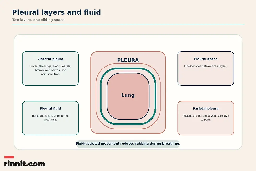
Structure | Location or characteristic |
Visceral pleura | Inner layer covering the lungs, blood vessels, bronchi and nerves; not pain-sensitive. |
Parietal pleura | Layer attached to the chest wall; pain-sensitive. |
Pleural space | Hollow area between the two pleural layers. |
Pleural fluid | Thin layer of fluid in the pleural space that helps the layers slide during breathing. |
What is pleural fluid?
Pleural fluid is the thin layer of fluid inside the pleural space. It allows the visceral and parietal pleura to slide against each other as the lungs move during breathing. [1]
Too much pleural fluid is called a pleural effusion. Blood or air can also enter the pleural space and put pressure on the lungs. [1]
What conditions affect the pleura?
Pleural disorders can involve inflammation, fluid, air, blood, injury or cancer. Six conditions are common.
Condition | Description |
Pleurisy | Inflammation in the pleura. |
Pleural effusion | Too much pleural fluid builds up in the pleural space. |
Hemothorax | Blood enters the pleural space, usually after an injury, and puts extra pressure on the lungs. |
Pneumothorax | Air enters the pleural space and puts pressure on the lungs. |
Hemopneumothorax | Blood and air enter the pleural space at the same time, usually after an injury. |
Pleural mesothelioma | Cancer that forms in the pleura. |
Pleurisy
Pleurisy is inflammation of the pleura. Because the parietal pleura is sensitive to pain, inflammation can be associated with breathing-related chest pain. [1]
Pleural effusion
A pleural effusion occurs when too much fluid builds up in the pleural space. A healthcare professional may use imaging or remove a fluid sample to evaluate the cause. [1]
Hemothorax
A hemothorax occurs when blood enters the pleural space, usually after an injury. The blood puts additional pressure on the lungs. [1]
Pneumothorax
A pneumothorax occurs when air enters the pleural space and puts pressure on the lungs. It may happen after an injury, and it can also occur with smoking or drastic air-pressure changes such as flying or scuba diving. [1]
Hemopneumothorax
A hemopneumothorax occurs when both blood and air enter the pleural space at the same time. It usually occurs after an injury. [1]
Pleural mesothelioma
Pleural mesothelioma is cancer that forms in the pleura. Possible treatments include radiation therapy, chemotherapy and immunotherapy, depending on the clinical situation.
What happens if the pleura is damaged?
Pleural damage may cause sharp pain that worsens when you breathe normally, take a deep breath or cough. You may also feel like you cannot get enough air into your lungs, a symptom called dyspnea. [1]
Damage can occur after a chest injury such as a fall, a contact-sport injury, a car accident or a puncture wound. If you have symptoms of severe pleural damage, call 911 or your local emergency number immediately. [1]

How are pleural conditions diagnosed?
Healthcare professionals may use imaging tests such as a chest X-ray, CT scan or ultrasound to evaluate a pleural condition. [1]
A thoracentesis uses a thin needle inserted between the ribs to remove a sample of pleural fluid. A thoracoscopy uses a small tube with a light and camera to look inside the chest and around the lungs. [1]
Test or procedure | What it may help evaluate |
Chest X-ray | Pleural and chest findings. |
CT scan | Detailed chest structures and possible pleural problems. |
Ultrasound | Pleural or fluid-related findings. |
Thoracentesis | A sample of pleural fluid. |
Thoracoscopy | The inside of the chest and areas around the lungs. |
The appropriate evaluation depends on symptoms, injury history and the suspected condition.
How are pleural conditions treated?
Treatment depends on the condition and its severity. Options include medication to treat an infection or relieve symptoms, oxygen therapy, draining fluid or air from the chest and surgery. Mechanical ventilation may also be used in some situations, and radiation therapy, chemotherapy or immunotherapy may be used when cancer affects the pleura. [1]
Treatment category | Use |
Medication | Treats an infection or relieves symptoms. |
Oxygen therapy | Supports breathing when clinically indicated. |
Drainage | Removes fluid or air from the chest. |
Surgery | May be used depending on the condition. |
Mechanical ventilation | A support option in some situations. |
Cancer treatment | Radiation therapy, chemotherapy or immunotherapy may be used for cancer. |
This list is educational and is not a treatment recommendation for an individual.
How can you help protect pleural health?
Pleural damage risk can be reduced by avoiding tobacco products, wearing a seatbelt in a car and wearing protective padding during contact sports. [1]
Prevention topic | Example |
Tobacco | Avoid smoking and vaping. |
Vehicle safety | Wear a seatbelt every time you are in a car. |
Sports protection | Wear protective padding during contact sports. |
Air pressure | Limit activities involving drastic pressure changes, such as flying or scuba diving. |
Infection prevention | Wash your hands regularly. |
Exposure reduction | Avoid asbestos. |
When should you seek medical help?
Call 911 or your local emergency number immediately for symptoms of severe pleural damage.
Contact a healthcare professional for sharp chest pain that worsens with breathing or coughing, dyspnea, a new chest injury or other concerning breathing symptoms. A sudden or severe breathing problem, rapidly worsening chest pain or symptoms after a significant chest injury need emergency care: call 911 or your local emergency number. This article cannot determine the urgency of an individual case.
Conclusion: A thin membrane with an important job
The pleura is a two-layered membrane that lines the chest wall and covers the lungs. Its pleural fluid helps the layers glide during breathing, while the membrane cushions the lungs and reduces rubbing against the chest wall. [1]
Inflammation, excess fluid, air, blood, injury and cancer can affect the pleura. Symptoms may include sharp breathing-related pain and dyspnea, and severe pleural-damage symptoms need immediate emergency help. Imaging, thoracentesis and thoracoscopy can help healthcare professionals evaluate pleural problems, and treatment depends on the condition. [1]
Use this guide to prepare questions for a qualified healthcare professional. Clear information is a starting point for better care, not a diagnosis. [1]
Frequently Asked Questions
What is the pleura?
The pleura is a thin, watery membrane that lines the chest wall and wraps around the lungs.
What is the function of the pleura?
The pleura cushions the lungs, helps prevent them from rubbing against the chest wall and lets the pleural layers slide during breathing.
What is pleural fluid?
Pleural fluid is a thin layer of fluid in the pleural space between the pleural layers. It helps them slide as you breathe.
What are the two layers of the pleura?
The visceral pleura is the inner layer covering the lungs. The parietal pleura attaches to the chest wall.
Which pleural layer feels pain?
The parietal pleura is sensitive to pain. The visceral pleura has no sensory nerves and is not sensitive to pain.
What is pleurisy?
Pleurisy is inflammation in the pleura.
What is a pleural effusion?
A pleural effusion is a buildup of too much fluid in the pleural space.
What is a pneumothorax?
A pneumothorax is air in the pleural space that puts pressure on the lungs. It may follow an injury or occur with smoking or drastic air-pressure changes.
How are pleural problems tested?
Healthcare professionals may use a chest X-ray, CT scan, ultrasound, thoracentesis or thoracoscopy.
When is pleural pain an emergency?
Call 911 or your local emergency number immediately for symptoms of severe pleural damage.
Related guides
References
Cleveland Clinic. Pleura: Location, Anatomy, Function, Diseases & Conditions. Cleveland Clinic. Updated July 22, 2024. https://my.clevelandclinic.org/health/body/pleura
Lakkadghatwala R, Wilson A, Sabhaney V, et al. Ultrasound guidance compared to anatomic landmark approach for thoracentesis: A systematic review and meta-analysis. Am J Emerg Med. 2025;97:159-164. PMID 40738083. https://doi.org/10.1016/j.ajem.2025.07.049
Brown SGA, Ball EL, Perrin K, et al. Conservative versus Interventional Treatment for Spontaneous Pneumothorax. N Engl J Med. 2020;382(5):405-415. PMID 31995686. https://doi.org/10.1056/NEJMoa1910775
Iyer NP, Reddy CB, Wahidi MM, et al. Indwelling Pleural Catheter versus Pleurodesis for Malignant Pleural Effusions. A Systematic Review and Meta-Analysis. Ann Am Thorac Soc. 2019;16(1):124-131. PMID 30272486. https://doi.org/10.1513/AnnalsATS.201807-495OC
Medical disclaimer
This article is for general educational purposes and is not a substitute for professional medical advice, diagnosis or treatment. Always seek the advice of a qualified healthcare professional with any questions about a medical condition or treatment.



Comments