Myelomeningocele: Causes, Diagnosis, Treatment and Long-Term Care

Updated: 2 days ago
Medically reviewed by Dr. Baraa Alnahhal, MD · Last reviewed: October 2026
Quick Answer: What is myelomeningocele?
Myelomeningocele is an open form of spina bifida that develops early in pregnancy when the neural tube does not close completely, leaving a fluid-filled sac on the baby's back containing part of the spinal cord and nerves. It can cause weakness, loss of sensation, bladder or bowel problems and paralysis. Treatment may include fetal or postnatal surgery and lifelong follow-up. [1]
What the research says
In the landmark randomized trial, repairing myelomeningocele before birth roughly halved the need for a shunt and improved motor outcomes, but increased risks for the pregnancy. The trial was stopped early for efficacy after 183 of a planned 200 women were recruited; 158 children were evaluated at 12 months. The first primary outcome (fetal or neonatal death or shunt placement by 12 months) occurred in 68% after prenatal surgery versus 98% after postnatal surgery (relative risk 0.70; 97.7% CI 0.58–0.84). Shunt placement was 40% versus 82%. Prenatal surgery also improved the mental development and motor composite score and ambulation at 30 months, but was associated with more preterm delivery and uterine dehiscence at delivery (Adzick et al., N Engl J Med, 2011). [2]
A later analysis of the same trial found milder orthopaedic needs after prenatal repair, though most orthopaedic conditions occurred at similar rates. Among all 183 patients (91 prenatal, 92 postnatal), rates of scoliosis, kyphosis, hip abnormality, clubfoot and tibial torsion did not differ. Leg-length discrepancy at 30 months was lower after prenatal repair (7% vs 16%; P = 0.047), and the proportion needing casting or bracing was also lower (78% vs 90%; P = 0.036) (Swarup et al., J Pediatr Orthop B, 2022). [3]

What is spina bifida?
Spina bifida is a broad term for congenital conditions in which the neural tube in the spinal area does not close completely. The neural tube develops into the brain and spinal cord during the first month of pregnancy. [1]
Myelomeningocele is one type of spina bifida and the most serious type. Other forms include meningocele and spina bifida occulta. [1]
Myelomeningocele compared with meningocele
Both conditions can cause a fluid-filled sac to protrude through an opening in a baby’s back. In meningocele, the sac contains fluid and the tissues covering the spinal cord, but the spinal cord itself is not in the sac. There is usually little or no nerve damage. [1]
In myelomeningocele, part of the spinal cord and nerves are in the sac and are damaged. This difference helps explain why myelomeningocele usually causes more significant neurological and mobility effects. [1]
Condition | What the sac contains | Typical neurological effect |
Myelomeningocele | Part of the spinal cord, nerves, meninges and cerebrospinal fluid. | Spinal-cord and nerve damage is common; weakness or loss of sensation may occur below the lesion. |
Meningocele | Fluid and coverings around the spinal cord, but not the spinal cord itself. | Usually little or no nerve damage. |
Spina bifida occulta | A smaller spinal closure difference without the open sac described above. | Effects vary and may be mild or absent. |

What happens in myelomeningocele?
During early fetal development, the two sides of the spine normally join to cover and protect the spinal cord, spinal nerves and meninges. In myelomeningocele, the neural tube does not close fully. [1]
The opening can occur anywhere along the spine. It most commonly develops in the lumbar or sacral areas of the low back. A fluid-filled sac protrudes through the opening and contains part of the spinal cord, nerves, meninges and cerebrospinal fluid. [1]
The spinal cord and nerves in the sac are damaged. The level of the lesion helps shape the effects: babies with a lower lesion typically have less severe symptoms than babies with a higher lesion.
In some cases, the sac opens before or during birth due to normal fetal movement. This is one reason prenatal planning and early postnatal care are important. [1]
What are the signs and symptoms?
A baby with myelomeningocele has a fluid-filled sac along the mid- to low-back. Healthcare professionals can often detect the condition during pregnancy with ultrasound. [1]
Because the spinal cord and nerves are affected, possible effects include:
Muscle weakness.
Loss of sensation below the lesion.
Decreased mobility.
Paralysis.
Loss of bladder control.
Loss of bowel control.
Other congenital, neurological or musculoskeletal issues may also occur. About 8 out of 10 infants with myelomeningocele have hydrocephalus, or fluid buildup in the brain. Associated conditions can include syringomyelia, a fluid-filled cyst within the spinal cord, and hip dislocation.
What causes myelomeningocele?
Healthcare professionals and researchers do not know one exact cause. A complex combination of genetic, nutritional and environmental factors appears to contribute. [1]
Low levels of folate or folic acid before and during early pregnancy appear to play a role. Folate supports fetal development of the brain and spinal cord. [1]
A risk factor does not mean that a person caused the condition. Most cases occur sporadically, and having a baby with myelomeningocele is not the pregnant person’s fault.
What are the risk factors?
Risk factors linked with myelomeningocele include folate or folic acid deficiency, a family history of neural tube defects, some anti-seizure medicines taken during pregnancy, poorly managed diabetes during pregnancy, obesity before pregnancy, and increased core body temperature early in pregnancy from a prolonged fever, sauna or hot-tub use. [1]
One child with a neural tube defect can increase the chance of a future pregnancy being affected. However, most babies with myelomeningocele are born to parents with no known family history of the condition.
Anyone who is pregnant or considering pregnancy should discuss prenatal vitamins, medication safety, diabetes management and other health concerns with a qualified healthcare professional. Do not stop an anti-seizure medicine or any other prescribed treatment without medical guidance.
Risk factor | What is known |
Low folate | Folate supports fetal brain and spinal-cord development; deficiency is linked with neural tube defects. |
Family history | A family history can increase risk, although many cases occur without one. |
Some anti-seizure medicines | Some are linked with a higher risk during pregnancy; medication decisions require professional guidance. |
Diabetes | Poorly managed diabetes during pregnancy is associated with higher risk. |
Obesity before pregnancy | Associated with increased risk of neural tube defects. |
Early-pregnancy hyperthermia | Prolonged fever or sauna or hot-tub exposure has been associated with a slightly increased risk. |
How is myelomeningocele diagnosed before birth?
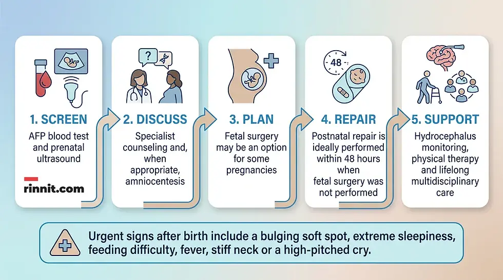
Healthcare professionals can usually, but not always, detect myelomeningocele during pregnancy. Prenatal evaluation may include blood screening, fetal ultrasound and amniocentesis. [1]
Alpha-fetoprotein screening
An AFP blood test is commonly measured during the 16th to 18th week of pregnancy. The AFP level is higher than normal in about 75% to 80% of pregnancies carrying a fetus with spina bifida. [1]
An elevated AFP result does not establish a diagnosis by itself. The healthcare professional may recommend additional testing, such as ultrasound, to evaluate the fetus more closely.
Fetal ultrasound
Fetal ultrasound is the most accurate method described for diagnosing myelomeningocele. Scans are commonly recommended during the first trimester, at 11 to 14 weeks, and the second trimester, at 18 to 22 weeks. Healthcare professionals can accurately diagnose myelomeningocele during the second-trimester scan. [1]
Amniocentesis
If an ultrasound confirms myelomeningocele, a healthcare professional may recommend amniocentesis to evaluate for rare associated genetic diseases. During amniocentesis, a needle removes a sample of fluid from the amniotic sac. [1]
Amniocentesis carries certain risks. The maternal-fetal medicine or prenatal team should explain the procedure, possible benefits and risks before a decision is made.
How is it diagnosed after birth?
If myelomeningocele was not identified during pregnancy, healthcare professionals may use MRI or CT after birth. The visible sac and newborn examination also help the care team understand the condition. [1]
Diagnosis is not the same as predicting every future ability. The level of the lesion, the condition of the spinal cord and nerves, whether the sac is open or closed and other associated conditions all affect the child’s needs.
How is myelomeningocele treated?
Treatment typically involves surgery to repair the opening in the baby’s spine. Repair may occur before birth through fetal surgery or shortly after birth through postnatal surgery. [1]
Babies often need evaluation for hydrocephalus. A ventriculoperitoneal, or VP, shunt may be placed to drain extra fluid from the brain. [1]
Most children need lifelong treatment for the effects of spinal-cord and nerve damage. Care may include physical therapy, support for mobility and treatment or prevention of infections such as meningitis or urinary tract infections.
Fetal surgery
Fetal surgery may be an option depending on the severity of the myelomeningocele and the health of the pregnant person. It may prevent symptoms from worsening, but it cannot correct spinal-cord or nerve damage that is already present. [1]
Risks associated with prenatal surgery include increased risk of preterm delivery and uterine dehiscence, which is thinning or tearing at the uterine incision during delivery. A specialized team should explain eligibility, potential benefits, risks and alternatives. [1]
Postnatal surgery
If fetal surgery was not performed, repair after birth should happen as soon as possible to reduce infection risk, ideally within the first 48 hours after birth.
The newborn team will protect the sac, monitor the baby and coordinate surgery and related evaluations. Parents can ask what will happen first, how the sac will be protected and when they can participate in care.
Which specialists may be involved?
Most children benefit from a multidisciplinary team. Specialists may include a neurologist, neurosurgeon, fetal surgeon, urologist, orthopaedist, dermatologist and physical therapist.
The team may expand as the child grows. Coordinated care can address neurological function, mobility, skin protection, bladder and bowel function, development, infections, learning and emotional well-being.
What complications can occur?
Myelomeningocele can cause paralysis, reduced mobility, bladder and bowel problems, infections, hydrocephalus and other complications, depending on the level of the spinal opening. [1]
Paralysis or loss of sensation below the lesion.
Decreased mobility from muscle weakness.
Scoliosis, contractures or hip dislocation.
Loss of bladder or bowel control.
Frequent urinary tract infections.
Meningitis.
Latex allergy.
Learning differences or delayed milestones.
Seizures.
Hydrocephalus.
Syringomyelia.
The level of the spinal opening and the condition of the spinal cord influence the pattern of effects. No two people with myelomeningocele are affected in exactly the same way.
What is the outlook?
The outlook depends on the initial condition of the spinal cord and nerves, how high or low the opening is, whether the sac is open or closed and whether surgery occurred before or after birth. [1]
People with spina bifida have a mortality rate of about 1% per year from ages 5 to 30, and a higher lesion is associated with greater risk of complications and mortality. These figures do not predict an individual child’s future. [1]
The effects of myelomeningocele can affect quality of life. Depression, anxiety and risk-taking behaviors are more common among people with spina bifida. Emotional support and developmental care are part of long-term health, not an afterthought. [1]
Can myelomeningocele be prevented?
Birth defects cannot be completely prevented, but risk can be reduced. Folate or folic acid is important for fetal development, so people who are pregnant or thinking about becoming pregnant should discuss prenatal vitamins and folate with a healthcare professional. The CDC recommends that anyone who could become pregnant take 400 micrograms of folic acid every day, starting at least one month before pregnancy; people who have had a pregnancy affected by a neural tube defect may be advised to take a higher dose.
Risk reduction also means reviewing medication safety before pregnancy, managing diabetes, discussing a family history of neural tube defects and seeking advice about fever or increased body temperature early in pregnancy.
Do not stop a prescribed medicine on your own. Some anti-seizure medicines are associated with increased risk, but untreated medical conditions can also be dangerous. A healthcare professional can help weigh the options.
How can people live well with myelomeningocele?
No two people with myelomeningocele are affected in the same way. It is not possible to predict every need from the prenatal diagnosis alone.
A child may benefit from regular care by a team experienced in spina bifida and myelomeningocele. The care plan can change as the child grows and develops.
When should I call my child’s healthcare provider?
Call the child’s healthcare provider if the child is late walking or crawling, has a bulging soft spot, is unusually irritable or extremely sleepy, has feeding difficulties, or has a fever, stiff neck, irritability or a high-pitched cry. [1]
A bulging soft spot, irritability, extreme sleepiness or feeding difficulty can be a sign of hydrocephalus. Fever, stiff neck, irritability or a high-pitched cry can be signs of meningitis. These symptoms require prompt medical attention.
What questions should you ask the myelomeningocele care team?
Families facing myelomeningocele can ask the care team about the opening's level, test results, fetal or after-birth surgery, hydrocephalus monitoring, long-term care needs and urgent warning signs.
Where is the opening, and what does its level mean?
Is the sac open or closed?
What does the ultrasound show about the spinal cord and brain?
Do we need AFP testing, amniocentesis, MRI or another evaluation?
Is fetal surgery an option, and what are its benefits and risks?
What will happen if repair occurs after birth?
How will the baby be protected before surgery?
How will the team monitor for hydrocephalus?
Could a VP shunt be needed?
What specialists should be involved after discharge?
How will bladder, bowel, mobility, skin and developmental needs be managed?
Which symptoms require an urgent call or emergency evaluation?
Where can our family find support groups and counseling?
Conclusion: Support begins with clear information
Myelomeningocele is an open form of spina bifida in which a fluid-filled sac contains part of the spinal cord, nerves, meninges and cerebrospinal fluid. It develops during the first four weeks of pregnancy and most often affects the low back. [1]
Prenatal ultrasound can often identify the condition. AFP screening and, when appropriate, amniocentesis may add information. Treatment usually involves fetal or postnatal surgery, with additional care for hydrocephalus, mobility, bladder and bowel function, infections, development and emotional health. [1]
The outlook varies widely. The level and condition of the spinal cord, the type of sac and the timing of repair all matter, but no single prenatal finding predicts every future need. A coordinated specialist team can help families make decisions and plan long-term support. [1]
Frequently Asked Questions
Is myelomeningocele the same as spina bifida?
Myelomeningocele is a type of spina bifida. Spina bifida is the broader term for congenital conditions in which the neural tube in the spinal area does not close completely.
What does the sac contain?
The sac contains part of the spinal cord, nerves, meninges and cerebrospinal fluid. The spinal cord and nerves inside the sac are damaged, which can cause weakness or loss of sensation below the lesion.
Where does myelomeningocele usually occur?
Myelomeningocele can occur anywhere along the spinal cord, but it most commonly affects the low back in the lumbar or sacral areas.
How is myelomeningocele detected during pregnancy?
Healthcare professionals may use AFP blood screening at 16 to 18 weeks, fetal ultrasound and sometimes amniocentesis. Ultrasound is the most accurate prenatal diagnostic method described.
Is a high AFP level proof of myelomeningocele?
No. AFP is a screening test. A higher-than-normal result may lead to an ultrasound or other evaluation; it does not establish the diagnosis by itself.
When can ultrasound diagnose myelomeningocele?
Healthcare professionals commonly perform first-trimester scans at 11 to 14 weeks and second-trimester scans at 18 to 22 weeks. Myelomeningocele can be accurately diagnosed during the second-trimester scan.
What is fetal surgery?
Fetal surgery repairs the spinal opening before birth. It may be an option depending on the severity of the condition and the pregnant person’s health. It may prevent symptoms from worsening but cannot repair existing nerve damage.
What happens if surgery occurs after birth?
Postnatal repair should occur as soon as possible to reduce infection risk, ideally within the first 48 hours. The newborn team protects the sac, monitors the baby and coordinates surgery and related evaluations.
Does every child with myelomeningocele need a shunt?
Not every child necessarily needs one, but hydrocephalus is common. About 8 out of 10 infants with myelomeningocele have fluid buildup in the brain, and a VP shunt may be used to drain extra fluid.
What complications can myelomeningocele cause?
Possible complications include weakness, paralysis, loss of sensation, mobility problems, bladder or bowel-control loss, UTIs, meningitis, hydrocephalus, scoliosis, contractures, hip dislocation, latex allergy, learning differences, delayed milestones and seizures.
Can myelomeningocele be prevented?
Myelomeningocele cannot be completely prevented, but risk can be reduced. Discuss folate or folic acid, prenatal vitamins, medication safety, diabetes management, family history and early-pregnancy fever or heat exposure with a healthcare professional.
Is myelomeningocele caused by something the pregnant person did?
No. Having a baby with myelomeningocele is not the pregnant person’s fault. The condition likely results from a complex combination of genetic, nutritional and environmental factors, and most cases occur sporadically.
What does the outlook depend on?
The outlook depends on the spinal-cord and nerve condition, the level of the opening, whether the sac is open or closed and whether repair occurred before or after birth. No two people are affected in exactly the same way.
What signs may indicate hydrocephalus in a baby?
A bulging soft spot, irritability, extreme sleepiness and feeding difficulties can be signs of hydrocephalus. Call the child’s healthcare provider promptly if these occur.
What signs may indicate meningitis?
Fever, stiff neck, irritability and a high-pitched cry can be signs of meningitis. Seek prompt medical attention.
Will my child need lifelong care?
Most children need lifelong management for effects of spinal-cord and nerve damage. The care team may include neurology, neurosurgery, urology, orthopaedics, physical therapy and other specialists.
Related guides
References
Cleveland Clinic. Myelomeningocele. Cleveland Clinic. Last updated April 19, 2022. https://my.clevelandclinic.org/health/diseases/22813-myelomeningocele
Adzick NS, Thom EA, Spong CY, et al. A randomized trial of prenatal versus postnatal repair of myelomeningocele. N Engl J Med. 2011;364(11):993-1004. PMID 21306277. https://doi.org/10.1056/NEJMoa1014379
Swarup I, Talwar D, Howell LJ, et al. Orthopaedic outcomes of prenatal versus postnatal repair of myelomeningocele. J Pediatr Orthop B. 2022;31(1):87-92. PMID 33165214. https://doi.org/10.1097/BPB.0000000000000827
Centers for Disease Control and Prevention. About Birth Defects. Centers for Disease Control and Prevention. Reviewed February 26, 2026. https://www.cdc.gov/birth-defects/about/index.html
Centers for Disease Control and Prevention. About Folic Acid. Centers for Disease Control and Prevention. Updated September 18, 2026. https://www.cdc.gov/folic-acid/about/index.html
Disclaimer
This article is for general educational purposes and is not a substitute for professional medical advice, diagnosis or treatment. Always seek the advice of a qualified healthcare professional with any questions about a medical condition or treatment.
Do not interpret an AFP result, ultrasound, amniocentesis finding or lesion level without the care team, and do not stop prescribed medication during pregnancy without professional guidance. Having a baby with myelomeningocele is not the pregnant person’s fault.



Comments