Lobular Carcinoma in Situ (LCIS): Symptoms, Causes, Diagnosis, and Treatment

Updated: 3 days ago
Medically reviewed by Dr. Baraa Alnahhal, MD · Last reviewed: October 2026
TL;DR: Lobular carcinoma in situ (LCIS) is a noncancerous cell change in the milk-producing glands of the breast. It causes no symptoms and is usually found by accident after a biopsy. Having LCIS raises your lifetime breast cancer risk to roughly 25%–30%, compared with about 13% for women in general, and affects both breasts. Management centers on closer screening, estrogen-blocking medicines taken for five years, or, for those at very high risk, preventive surgery.
Quick Answer: What is lobular carcinoma in situ?
Lobular carcinoma in situ (LCIS) is a growth of abnormal-looking cells inside a milk-producing lobule of the breast. It is not cancer and does not invade nearby tissue, but it marks an increased breast cancer risk. LCIS causes no symptoms and is usually found during a biopsy. Management includes closer screening, estrogen-blocking medicine and sometimes preventive mastectomy.
What the research says
Breast cancer risk stays raised for years after an LCIS diagnosis. A meta-analysis of 70 studies (47,671 people) estimated that breast cancer developed in 9.3% of people within 5 years of an LCIS diagnosis and in 11.8% within 10 years (Baker et al., Breast, 2024 [3]).
Cancer is rarely found next to LCIS seen on a needle biopsy. Across 16 high-quality studies, the pooled risk that classic LCIS on a core needle biopsy was upgraded to cancer after surgery was 5.8%, and 3.5% for invasive cancer (Shehata et al., J Am Coll Radiol, 2020 [4]).
What Is Lobular Carcinoma in Situ?
Lobular carcinoma in situ (LCIS) is a growth of cells inside a milk gland — also called a lobule — in the breast. The cells change in ways that make them look different from typical cells. Despite the word "carcinoma" in its name, LCIS is not cancer. Instead, it is a sign that a person's risk of developing breast cancer in the future is higher than average.
The word "in situ" means the changed cells stay in place. They remain inside the lobule and do not extend into or invade nearby breast tissue.
LCIS is unusual in how it is discovered. It usually does not show up on a mammogram and cannot be felt during a breast exam. Most of the time, it is found by accident after a procedure to remove some breast tissue, such as a breast biopsy or a lumpectomy.
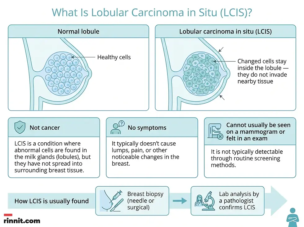
One important fact shapes how LCIS is managed: it increases the risk of breast cancer in both breasts, not just the breast where the cells were found. Because of this, a diagnosis usually leads to increased breast cancer screening. You may also want to consider treatments that reduce the risk of developing invasive breast cancer.
How Breast Tissue Is Organized
Understanding LCIS is easier with a quick look at how breast tissue is built. Each breast contains 15 to 20 lobes of glandular tissue, arranged like the petals of a daisy. Each lobe is divided into smaller units called lobules, which are the milk-producing glands. Small tubes called ducts carry milk from the lobules to a reservoir just beneath the nipple. LCIS begins in these milk-producing lobules.
Breast structure | What it does |
Lobes (15 to 20 per breast) | Main units of glandular tissue, arranged like daisy petals |
Lobules | Smaller units inside lobes that produce milk for breastfeeding |
Ducts | Small tubes that conduct milk from the lobules to the nipple area |
Milk reservoir | Lies just beneath the nipple; receives milk from the ducts |
What are the symptoms of lobular carcinoma in situ (LCIS)?
LCIS does not cause any signs or symptoms. There is no lump to feel, no pain, and no visible change on the skin.
Because of this, LCIS is usually found after a procedure to remove some breast tissue — such as a breast biopsy performed for another reason, or a lumpectomy.
Symptom-related question | What LCIS does |
Does it cause a lump? | No — LCIS does not produce a lump |
Does it cause pain or nipple discharge? | No — LCIS has no symptoms |
Can you feel it during a breast exam? | Usually not |
Can it be seen on a mammogram? | Usually not |
How is it typically found? | By accident during a breast biopsy or after a lumpectomy |
When should you see a doctor about lobular carcinoma in situ (LCIS)?
Lobular carcinoma in situ (LCIS) causes no symptoms, but it is wise to see a healthcare professional if you notice any change in your breasts. Watch for these changes:
Warning sign | What to look for |
A lump | Any new lump or firm area in the breast or underarm |
Skin changes | An area of puckered or otherwise unusual skin |
Thickened skin | A thickened region under the skin |
Nipple discharge | Any discharge from the nipple |
When you make an appointment, ask two important questions: when you should consider breast cancer screening, and how often that screening should be repeated. Most healthcare professionals recommend routine breast cancer screening beginning in your 40s.
What Causes Lobular Carcinoma in Situ?
The exact cause of lobular carcinoma in situ (LCIS) is not clear, though researchers understand how it begins. Here is what is understood about how it begins.
LCIS starts when cells in a milk-producing gland of the breast — a lobule — develop changes in their DNA. These DNA changes cause the cells to look different from healthy cells. The changed cells remain in the lobule and do not extend into, or invade, nearby breast tissue. This is why LCIS is not considered cancer.
However, having LCIS means you are more likely to develop invasive breast cancer later. The risk applies to both breasts.
Your Risk Numbers
Scenario | Estimated lifetime risk of breast cancer |
Women in general | About 13% |
Women diagnosed with LCIS | About 25% to 30% |
Your individual risk depends on many factors beyond the LCIS finding itself. Talk to your healthcare team to understand your personal risk.
How is lobular carcinoma in situ (LCIS) diagnosed?
LCIS is most often diagnosed during a breast biopsy — a procedure that removes a sample of breast tissue for testing. Because LCIS often cannot be detected on a mammogram or felt during a breast exam, biopsy is usually the only way it is found.

Two types of breast biopsy may be used:
Biopsy type | How it works |
Core needle biopsy | A healthcare professional uses a thin, hollow needle to remove several tiny tissue samples from the breast. Imaging tests such as ultrasound or MRI are often used to help guide the needle. |
Surgical biopsy | A surgeon performs an operation to remove the tissue sample for testing. |
The tissue removed during a biopsy is sent to a laboratory. There, doctors who specialize in analyzing blood and body tissues — called pathologists — closely examine the cells. They can determine whether LCIS is present.
What are the treatment options for lobular carcinoma in situ (LCIS)?
Treatment for LCIS often involves close monitoring for signs of cancer. Medicines or surgery may also be used to lower the risk of breast cancer. Your healthcare team considers many factors when creating a treatment plan, including your overall health and your preferences.

Careful Monitoring for Breast Cancer
If you have been diagnosed with LCIS, you may need more frequent exams to look for signs of breast cancer. A monitoring plan may include several layers of screening:
Monitoring step | Frequency |
Mammograms | Annually |
Breast exams by a healthcare professional | Every 6 to 12 months |
Breast self-exams | Monthly — to develop breast awareness and detect changes |
Additional screening tests | Based on your other risk factors |
The additional screening tests may include breast MRI, molecular breast imaging, and other supplemental breast imaging tests. Which test is best for you may depend on your risk factors for breast cancer, your family history of breast cancer, whether you have DNA changes that increase breast cancer risk, and whether your mammogram images show that you have dense breasts.
Medicine to Lower the Risk of Breast Cancer
Most breast cancers use estrogen and other hormones to help the cancer grow. Medicines that block the hormone estrogen in the body can lower the risk of breast cancer — blocking estrogen helps stop cancer from forming.
Four hormone-blocking medicines are used to lower breast cancer risk:
Medicine | Notes |
Tamoxifen | Sometimes taken at a lower dose every other day |
Raloxifene (Evista) | Taken daily |
Anastrozole (Arimidex) | Taken daily |
Exemestane (Aromasin) | Taken daily |
These medicines are typically taken daily for five years to reduce the risk of breast cancer. Raloxifene, anastrozole and exemestane are used only after menopause. Tamoxifen and raloxifene carry FDA boxed warnings for blood clots and stroke, and tamoxifen also for uterine cancer, so discuss these risks with your doctor. Talk with your healthcare team about which medicine is best for you.
Surgery to Lower the Risk of Breast Cancer
If your risk of breast cancer is very high, your healthcare team might recommend surgery to lower that risk. Risk may be considered high if you have a strong family history of breast cancer, or if DNA changes that increase the risk of breast cancer run in your family.
One operation that can lower the risk of breast cancer is surgery to remove one or both breasts. This procedure is called a risk-reducing mastectomy or a prophylactic mastectomy.
Treatment for Pleomorphic Lobular Carcinoma in Situ
Surgery is often recommended for a specific type of LCIS called pleomorphic lobular carcinoma in situ. This type is thought to carry a greater risk of breast cancer than other forms.
Pleomorphic LCIS is not common. Unlike other kinds of LCIS, it can be seen on a mammogram.
If a biopsy confirms pleomorphic LCIS, your healthcare team will likely recommend surgery. The two options are:
Surgery type | What it involves |
Lumpectomy | An operation to remove the area of concerning cells |
Mastectomy | An operation to remove nearly all of the breast tissue |
To decide which treatment is best for you, your healthcare team considers how much of your breast tissue needs to be removed, the results of your mammogram, whether you have a strong family history of cancer, and your age.
How can people cope with lobular carcinoma in situ (LCIS)?
Although LCIS is not cancer, a diagnosis can cause worry about the increased risk of a future breast cancer. Coping often means finding a long-term way to manage fear and worry. Three approaches can help.
Learn enough to make decisions about your care. Ask your healthcare team questions about your LCIS and what it means for your breast cancer risk. Use this information to research your treatment options. Look to reputable sources of information, such as the U.S. National Cancer Institute, to find out more. This can make you feel more confident as you make choices about your care.
Go to all of your screening appointments. You may feel anxious before breast cancer screening exams. Do not let that stop you from going. Plan ahead and expect that you will be anxious. To cope, spend time doing things you enjoy in the days before your appointment — time with friends or family, or quiet time for yourself.
Control what you can about your health. Make healthy lifestyle changes so you can feel your best. Choose a healthy diet that focuses on fruits, vegetables, and whole grains. Try to be active for 30 minutes most days of the week. Get enough sleep each night so that you wake feeling rested.
How should you prepare for an LCIS appointment?
Make an appointment with a doctor or other healthcare professional if you notice a lump or any other unusual change in your breasts.
What You Can Do
Before your appointment, gather and organize the following information:
Preparation step | What to do |
Record your symptoms | Write down any symptoms you are experiencing and for how long. If you have a lump, note when you first noticed it and whether it seems to have grown |
Document family history | Note which family members have had cancer, how each is related to you, the type of cancer, the age at diagnosis, and whether each person survived |
List your medications | Make a list of all medicines, vitamins, or supplements you are taking. Tell your provider about any hormone replacement therapy, current or past |
Bring support | Consider taking a family member or friend along — someone may remember something you missed |
Write down questions | Prepare a prioritized list of questions to make the most of your limited appointment time |
Questions to Ask Your Healthcare Professional
Some basic questions worth asking about LCIS include:
How much does lobular carcinoma in situ increase my risk of breast cancer?
Do I have any additional risk factors for breast cancer?
How often should I be screened for breast cancer?
What types of screening technology will be most effective in my case?
Am I a candidate for medicines that reduce the risk of breast cancer?
What are the possible side effects or complications of these medicines?
Which medicine do you recommend for me, and why?
How will you monitor me for treatment side effects?
Am I a candidate for preventive surgery?
In general, how effective is the treatment you're recommending in women with a diagnosis similar to mine?
What lifestyle changes can help reduce my risk of cancer?
Do I need a second opinion?
Should I see a genetic counselor?
What to Expect From Your Doctor
Be prepared to answer questions about your symptoms and your health. Your doctor may ask:
Do you have a breast lump that you can feel?
When did you first notice this lump?
Has the lump grown or changed over time?
Have you noticed any other unusual changes in your breast, such as discharge, swelling, or pain?
Have you gone through menopause?
Are you using or have you used any medicines or supplements to relieve the symptoms of menopause?
Have you been diagnosed with any previous breast conditions, including noncancerous conditions?
Have you been diagnosed with any other medical conditions?
Do you have any family history of breast cancer?
Have you or your close female relatives ever been tested for BRCA gene changes?
Have you ever had radiation therapy?
What is your typical daily diet, including alcohol intake?
Are you physically active?
Conclusion
Lobular carcinoma in situ can feel alarming, but it is important to remember what LCIS actually is: a noncancerous cell change that raises your future breast cancer risk — not cancer itself. It causes no symptoms, cannot usually be felt or seen on imaging, and is most often found by accident during a biopsy. The lifetime breast cancer risk for women with LCIS is thought to be about 25% to 30%, roughly double the average of 13%, and that risk applies to both breasts.
The good news is that you have clear, proven options for managing that risk: annual mammograms and regular clinical exams, estrogen-blocking medicines taken for five years, and — for those with very high risk — preventive surgery.
Ready to take the next step? If you have noticed any change in your breasts, or if you have questions about screening as you move into your 40s, book an appointment with your healthcare professional. Getting a personalized risk assessment and a monitoring plan in place is the best way to stay ahead of your health.
FAQ
What is lobular carcinoma in situ (LCIS)?
LCIS is a growth of cells inside a milk-producing gland (lobule) of the breast. The cells look different from normal cells, but LCIS is not cancer. The changed cells stay in the lobule and do not invade nearby tissue. It is a marker of increased breast cancer risk.
Is lobular carcinoma in situ cancer?
No. LCIS is not cancer. The abnormal cells remain in the lobule and do not extend into or invade nearby breast tissue. However, having LCIS raises the risk of developing invasive breast cancer in the future, in both breasts.
What are the symptoms of lobular carcinoma in situ?
LCIS does not cause any signs or symptoms. It usually cannot be felt during a breast exam or seen on a mammogram. It is most often found by accident after a breast biopsy or a lumpectomy.
How much does LCIS increase breast cancer risk?
The lifetime breast cancer risk for women diagnosed with LCIS is thought to be approximately 25% to 30%, compared with about 13% for women in general. The increased risk applies to both breasts. Individual risk depends on many factors.
How is lobular carcinoma in situ diagnosed?
LCIS is most often diagnosed during a breast biopsy, a procedure that removes a sample of breast tissue for testing. Tissue may be taken with a core needle biopsy (sometimes guided by ultrasound or MRI) or a surgical biopsy. Pathologists examine the cells in a lab to confirm LCIS.
What are the treatment options for LCIS?
Management usually starts with close monitoring: annual mammograms, breast exams every 6 to 12 months, and monthly self-exams. Estrogen-blocking medicines such as tamoxifen, raloxifene, anastrozole, or exemestane — typically taken daily for five years — can lower breast cancer risk. For very high-risk cases, preventive mastectomy is an option.
Can LCIS be seen on a mammogram?
Usually not. Most LCIS cannot be detected on a mammogram or felt during a breast exam. The exception is a rare type called pleomorphic lobular carcinoma in situ, which can be seen on a mammogram and is usually treated with surgery.
Does LCIS have to be treated with surgery?
Not always. For most LCIS, treatment involves close monitoring and possibly estrogen-blocking medicine. Surgery is considered mainly for pleomorphic LCIS (lumpectomy or mastectomy) or for people with very high risk due to family history or inherited DNA changes (preventive mastectomy).
What medicines lower breast cancer risk in LCIS?
Hormone-blocking medicines that block estrogen can lower breast cancer risk, because most breast cancers use estrogen to grow. The medicines used are tamoxifen, raloxifene (Evista), anastrozole (Arimidex), and exemestane (Aromasin), typically taken daily for five years.
When should I see a doctor about my breasts?
Make an appointment if you notice a change in your breasts, such as a lump, puckered or unusual skin, a thickened region under the skin, or nipple discharge. Most healthcare professionals recommend routine breast cancer screening beginning in your 40s.
External Resources
Related Reading
Breast Biopsy: Types, What to Expect, Recovery and Results explains the procedure used to confirm or rule out breast cancer.
References
Mayo Clinic Staff. Lobular carcinoma in situ (LCIS) — Symptoms and causes. Mayo Clinic. July 28, 2025. https://www.mayoclinic.org/diseases-conditions/lobular-carcinoma-in-situ/symptoms-causes/syc-20374529
Mayo Clinic Staff. Lobular carcinoma in situ (LCIS) — Diagnosis & treatment. Mayo Clinic. July 28, 2025. https://www.mayoclinic.org/diseases-conditions/lobular-carcinoma-in-situ/diagnosis-treatment/drc-20374535
Baker J, Noguchi N, Marinovich ML, et al. Atypical ductal or lobular hyperplasia, lobular carcinoma in-situ, flat epithelial atypia, and future risk of developing breast cancer: systematic review and meta-analysis. Breast. 2024;78:103807. PMID: 39270543. DOI: https://doi.org/10.1016/j.breast.2024.103807
Shehata MN, Rahbar H, Flanagan MR, et al. Risk for upgrade to malignancy after breast core needle biopsy diagnosis of lobular neoplasia: a systematic review and meta-analysis. J Am Coll Radiol. 2020;17(10):1207-1219. PMID: 32861602. DOI: https://doi.org/10.1016/j.jacr.2020.07.036
Medical disclaimer
This article is for informational purposes only and is not a substitute for professional medical advice, diagnosis, or treatment. The clinical information in this guide reflects source material last reviewed in July 2025.



Comments