Dysplastic Nevus (Atypical Moles): What They Look Like, Your Melanoma Risk and What to Do Next

Updated: 3 days ago
Medically reviewed by Dr. Baraa Alnahhal, MD · Last reviewed: November 2026
Quick Answer
A dysplastic nevus is an atypical mole with irregular borders, mixed colors and a size larger than a pencil eraser [1]. Atypical moles are benign, but people with 10 or more are 12 times more likely to develop melanoma [1]. A dermatologist diagnoses them, monthly ABCDE self-checks help, and most need no treatment, though early detection is lifesaving [1].
What the research says
The number of atypical moles tracks closely with melanoma risk. A meta-analysis of 46 observational studies found that having 5 atypical nevi versus none was linked to a pooled relative risk of 6.36 (95% CI 3.80–10.33) for melanoma, while 101–120 common nevi versus fewer than 15 carried a pooled relative risk of 6.89 (95% CI 4.63–10.25); case-control studies, especially hospital-based ones, gave lower and more precise estimates than cohort studies (PubMed: Gandini et al., Eur J Cancer, 2005 [2]).
Mole count is one of the best-supported melanoma risk factors. An umbrella review of 44 papers covering 85 associations between non-genetic factors and skin cancers found that only 13 had a high level of credibility; common and atypical nevi were among them for melanoma, alongside freckle density, eye color, hair color, skin type, sunburns and a history of melanoma. The authors cautioned that most meta-analyses in the field showed large heterogeneity or small-study bias (PubMed: Belbasis et al., J Dermatol Sci, 2016 [3]).
What Is a Dysplastic Nevus?
A dysplastic nevus is a mole that looks different from most moles. The mole may have irregular borders, be a mix of colors and appear larger than other moles [1].
Atypical moles are benign (not cancerous) [1]. However, having a lot of atypical moles increases your risk of melanoma, a life-threatening skin cancer [1] [2]. Atypical moles may also progress over time to become cancerous [1].

What are moles, exactly?
Specialized cells called melanocytes give your skin its color. When these skin cells clump together, they form moles (nevi) [1].
Most people have harmless moles known as common moles. It's normal for adults with light skin to have up to 40 common moles [1]. Common and atypical moles tend to first appear during childhood and adolescence [1].
Breaking down the medical term
You may better understand this condition when you break down the meaning of each word.
Term | Meaning |
Dysplastic (dysplasia) | The presence of unusual (abnormal) cells in tissue. Dysplastic tissue isn't cancerous, but it has the potential to develop into cancer. |
Nevus | The medical term for a mole, a common type of skin growth. |
Nevi | Multiple moles. Your provider may use "dysplastic nevi" if you have multiple atypical moles. |
How common are atypical moles?
Atypical moles are common. Experts estimate that 1 in 10 Americans have at least one atypical mole [1].
Do atypical moles cause skin cancer?
Having atypical moles increases your risk of melanoma. The more atypical moles you have, the greater the cancer risk [1] [2]. People who have 10 or more atypical moles are 12 times more likely to develop melanoma [1]. Still, most people with atypical moles don't develop melanoma [1].
Experts estimate that 1 in 4 cases of melanoma arises from a dysplastic nevus or atypical mole [1].
Other names for dysplastic nevus
Your healthcare provider may use these terms to refer to dysplastic nevus: atypical mole and Clark's nevus; congenital nevi and Spitz nevi are different types of moles [1].
What Do Atypical Moles Look Like?
An atypical mole (dysplastic nevus) may have some or all of these features:
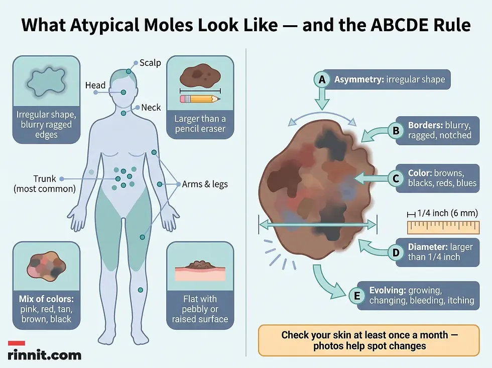
Where do atypical moles appear?
Atypical moles can appear anywhere on your body. They most often appear on your trunk but can also show up on your extremities (arms and legs), head, neck and scalp [1].
What Causes Atypical Moles?
Experts aren't sure why some people develop dysplastic nevi. A combination of genetics (family history) and environmental factors like ultraviolet (UV) light exposure may play a role [1].
The sun is a natural source of UV light. Artificial sources include certain halogen, incandescent and fluorescent lights and some lasers, mercury vapor lighting used in large venues like stadiums and gyms, and tanning beds and sunlamps [1].
Who is at risk?
Atypical moles can affect people of all ages, sexes and skin colors [1]. Having dark skin doesn't protect you from atypical moles or skin cancer, but your risk is reduced [1].
People with these risk factors are more likely to develop atypical moles:
Risk factor | Details |
Skin and hair type | Fair skin, freckles, light eyes and hair |
Family history | Family history of atypical moles, skin cancer or melanoma |
Sun history | Excessive sun exposure, sunburns or an inability to tan |
UV sensitivity | Sensitivity to UV light (photosensitivity) |
Possible complications
In addition to melanoma, having a lot of atypical moles may put you at risk for ocular melanoma — a type of cancer that develops in your eyes [1]. You may need more frequent eye exams [1].
How Are Dysplastic Nevi Diagnosed?
A dermatologist diagnoses and treats skin diseases like skin cancer. Seeing a skin cancer specialist is important when you have atypical moles. Your healthcare provider may perform regular skin examinations once or twice a year to check for changes to moles (or every three to six months if you have a family history of melanoma) [1].
Your provider may also use specialized tools:
Test | What it involves |
Skin examination | In-office check of moles, once or twice a year (every 3–6 months with family history of melanoma) |
Dermoscopy | A magnifying device called a dermatoscope sees abnormalities in moles invisible to the naked eye |
Mole mapping | A special camera with a dermatoscopic lens creates a computerized map of your skin and moles |
How to check your moles at home
You should perform skin self-exams regularly to look for changes to moles or skin. Healthcare providers recommend checking your skin at least once a month if you have atypical moles or other skin cancer risk factors [1]. You can also take photographs to help detect changes from month to month [1].
You can use the ABCDE rule while checking moles for signs of skin cancer:
Letter | Warning sign |
A — Asymmetry | Asymmetrical or irregular shape |
B — Borders | Borders that are blurry, ragged or notched |
C — Color | Colors that include a mix of shades like browns, blacks, reds or blues |
D — Diameter | Diameter larger than a pencil eraser (1/4 of an inch) |
E — Evolving | Changes over time: moles that grow in size, change shape or color, bleed or itch |
Contact a healthcare provider if you notice anything unusual, even if you think it's nothing.

How Are Dysplastic Nevi Treated?
An atypical mole doesn't need further treatment unless a skin biopsy indicates cancer or pre-cancerous changes [1].
Your healthcare provider may use the terms atypical melanocytic hyperplasia or moderately to severely dysplastic to describe biopsy results. Such results indicate a higher risk of cancer. Your provider will then treat the mole with further surgery.
Treatments for atypical moles involve surgically removing the mole and a small border of surrounding skin [1]. If the biopsy shows cancer (melanoma) associated with the atypical mole, your provider may perform a more extensive surgical removal or Mohs surgery, which helps preserve surrounding healthy tissue [1].
What Is the Outlook?
Most people with atypical moles (as well as common moles) don't develop skin cancer [1]. Still, it's important to examine your skin regularly for any unusual changes. Early detection and treatment of melanoma can be lifesaving.
Related conditions: atypical mole syndrome and FAMMM
Atypical mole syndrome describes people who have large numbers of moles and an increased risk of melanoma. Most of the moles are common, but some are atypical moles. Some moles may be larger than a third of an inch in diameter [1].
A different condition, familial atypical multiple mole melanoma (FAMMM) syndrome, affects members of the same family. An inherited gene mutation increases your risk of having atypical moles and melanoma. People with FAMMM syndrome are 25 times more likely to develop melanoma [1].
Can You Prevent Atypical Moles?
There isn't anything you can do to keep from having atypical moles. If you have them, you should take steps to protect your skin from sun damage and never use tanning beds [1].
It's also worth knowing that while having atypical moles increases your risk of melanoma, removing benign normal moles doesn't lower this risk [1].
Regular skin self-exams can help you quickly identify changes that may indicate a problem. Your healthcare provider can show you how to do these exams and tell you what changes to look for. Your provider may also recommend more frequent in-office skin exams, as well as eye exams. Contact your provider any time you notice changes to your skin.
Key Takeaways
Atypical moles are common, benign skin growths that simply look different from the average mole. The important nuance is risk, not certainty: most people with atypical moles never develop melanoma, yet having ten or more raises the odds twelvefold, and roughly one in four melanomas starts in an atypical mole. That makes consistent monitoring your best protection. Monthly ABCDE self-exams with photographs, regular dermatologist visits, and prompt reporting of any evolving mole give you the earliest possible warning — and early detection of melanoma is lifesaving. Protect your skin from the sun, skip tanning beds entirely, and never hesitate to contact your provider about a mole that looks or feels unusual.
Take the Next Step
If you have atypical moles, schedule a baseline skin exam with a dermatologist and ask how often you should return, and whether you should add more frequent eye exams to your schedule.
Frequently Asked Questions
1. What is a dysplastic nevus?
A dysplastic nevus is a mole that looks different from most moles. It may have irregular borders, be a mix of colors and appear larger than other moles. Atypical moles are benign (not cancerous), but having many increases melanoma risk and they may progress over time to become cancerous.
2. Are atypical moles cancerous?
No. Atypical moles are benign (not cancerous). However, having a lot of atypical moles increases your risk of melanoma, a life-threatening skin cancer, and the moles may progress over time to become cancerous.
3. How common are atypical moles?
Atypical moles are common. Experts estimate that 1 in 10 Americans have at least one atypical mole.
4. Do atypical moles cause melanoma?
Having atypical moles increases your risk of melanoma — the more you have, the greater the risk. People with 10 or more atypical moles are 12 times more likely to develop melanoma. Experts estimate 1 in 4 melanoma cases arises from a dysplastic nevus. Still, most people with atypical moles never develop melanoma.
5. What do atypical moles look like?
An atypical mole may be flat with a pebbly or slightly raised surface, have an irregular shape with blurry or ragged edges, be larger than a pencil eraser, and show a mix of colors including pink, red, tan, brown and black.
6. Where do atypical moles appear?
Atypical moles can appear anywhere on your body. They most often appear on the trunk but can also show up on the arms, legs, head, neck and scalp.
7. How are atypical moles diagnosed?
A dermatologist performs regular skin examinations (once or twice a year, or every 3–6 months with a melanoma family history), dermoscopy with a dermatoscope, and mole mapping using a special camera that creates a computerized map of your skin and moles.
8. What is the ABCDE rule for moles?
The ABCDE rule checks moles for Asymmetry (irregular shape), Borders (blurry, ragged or notched), Color (a mix of browns, blacks, reds or blues), Diameter (larger than a pencil eraser, 1/4 inch) and Evolving changes (growing, changing shape or color, bleeding or itching).
9. How are dysplastic nevi treated?
An atypical mole doesn't need treatment unless a skin biopsy indicates cancer or pre-cancerous changes. Treatment involves surgically removing the mole and a small border of surrounding skin. If melanoma is found, more extensive removal or Mohs surgery may be performed.
10. Can you prevent atypical moles?
There isn't anything you can do to keep from having atypical moles. If you have them, protect your skin from sun damage and never use tanning beds. Removing benign normal moles doesn't lower melanoma risk.
11. What is atypical mole syndrome?
Atypical mole syndrome means having large numbers of moles — most common, some atypical — with an increased melanoma risk; some moles may be larger than a third of an inch. FAMMM syndrome is an inherited condition in family members that makes melanoma 25 times more likely.
Related Reading
Learn more in our guide to Skin Biopsy: Types, What to Expect, Results and Aftercare.
Skin Self-Exam: How to Check Your Skin and the ABCDEs of Melanoma explains how to check your own skin monthly using the ABCDEs of melanoma.
References
Cleveland Clinic. Dysplastic Nevus (Atypical Moles). Cleveland Clinic. Mar. 21, 2023. https://my.clevelandclinic.org/health/diseases/24841-dysplastic-nevus-atypical-moles
Gandini S, Sera F, Cattaruzza MS, et al. Meta-analysis of risk factors for cutaneous melanoma: I. Common and atypical naevi. Eur J Cancer. 2005;41(1):28-44. doi:10.1016/j.ejca.2004.10.015. PMID: 15617989.
Belbasis L, Stefanaki I, Stratigos AJ, Evangelou E. Non-genetic risk factors for cutaneous melanoma and keratinocyte skin cancers: An umbrella review of meta-analyses. J Dermatol Sci. 2016;84(3):330-339. doi:10.1016/j.jdermsci.2016.09.003. PMID: 27663092.
Medical Disclaimer
This article is for informational purposes only and does not constitute medical advice. Always seek the advice of your physician or other qualified health provider with any questions you may have regarding a medical condition. Never disregard professional medical advice or delay in seeking it because of something you have read here.



Comments