Corneal Dystrophy: Symptoms, Causes, Types, Diagnosis, and Treatment

Updated: 2 days ago
Medically reviewed by Dr. Baraa Alnahhal, MD · Last reviewed: October 2026
Quick Answer
Corneal dystrophy is a group of more than 20 lifelong, genetic eye diseases that damage the cornea, the clear front layer of the eye. They are present from birth, usually affect both eyes and worsen gradually. They cannot be prevented, but most people keep functional vision with treatments ranging from eyedrops to laser removal or cornea transplant. [1]
What the research says
Experts classify corneal dystrophies into four groups. The International Classification of Corneal Dystrophies (IC3D, edition 2) groups them as epithelial and subepithelial, epithelial-stromal (TGFBI), stromal, and endothelial dystrophies, based on updated clinical, histologic, and genetic evidence (Weiss et al., Cornea, 2015) [2].
Fuchs dystrophy is the most common endothelial type. A review describes Fuchs endothelial corneal dystrophy as the most common primary corneal endothelial dystrophy and the leading reason for corneal transplantation worldwide; it typically appears around the fifth decade of life, is more common in women, and involves progressive loss of endothelial cells (Ong Tone et al., Progress in Retinal and Eye Research, 2021) [3].
Laser treatment can help, but problems often return. A review of phototherapeutic keratectomy (PTK) found it improves corneal clarity and smooths the surface, but the original problem and corneal haze often recur over the long term; repeat ablation is possible without affecting a future transplant, which makes PTK especially valuable where donor tissue is scarce (Nagpal et al., Survey of Ophthalmology, 2020) [4].
What Is Corneal Dystrophy?
Corneal dystrophy is an umbrella term for over 20 different diseases that affect the cornea of your eye. These diseases are present from birth and are lifelong, but they are usually possible to treat or manage. [1]
Dystrophy-type corneal diseases cause tissue changes or damage that disrupt how light passes through your corneas. If the disruptions are severe enough, they can decrease how well you see. [1]
These conditions are genetic. They happen because of DNA changes. Some changes are inherited from your biological parents. Others happen spontaneously. A few of these diseases happen for reasons experts do not fully understand yet. [1]
Key idea: Think of your cornea as commercially made glass engineered to be clear, flawless, and dome-shaped. Corneal dystrophies add unwanted tissue buildup, like looking through flaw-riddled blown glass that is harder to see through. [1]
Key Facts at a Glance
Fact | Detail |
Definition | Umbrella term for over 20 genetic corneal diseases |
Present from birth | Yes: lifelong conditions |
Cause | DNA changes (inherited or spontaneous) |
Eyes affected | Usually both eyes |
Preventable | No |
Causes total blindness | Rare: severe vision loss possible, not total blindness |
Most common type | Fuchs endothelial corneal dystrophy (about 39% of U.S. corneal transplants) |
How Are the Types of Corneal Dystrophy Classified?
The types of corneal dystrophies are grouped by the layer(s) of the cornea in which they happen. There are four main families. [1]
Type Family | Conditions Included | Typical Onset |
Epithelial and subepithelial | Epithelial basement membrane, epithelial recurrent erosion (3 subtypes), gelatinous drop-like, Lisch epithelial, Meesmann, subepithelial mucinous | Early childhood (mostly); basement membrane type usually adulthood; gelatinous drop-like any time before age 20 |
Epithelial-stromal | Granular (type 1 and type 2), lattice (multiple subtypes), Reis-Bückler's, Thiel-Behnke | Childhood; lattice type 1 as late as age 20 |
Stromal | Macular, Schnyder crystalline, congenital hereditary stromal, fleck, posterior amorphous, Pre-Descemet, central cloudy of François | Childhood mostly; Schnyder as late as age 30; Pre-Descemet usually after age 30 |
Endothelial | Fuchs endothelial, posterior polymorphous, congenital hereditary endothelial, X-linked endothelial | Varies |
Which Type Is Most Common?
Of all the corneal dystrophies, Fuchs endothelial corneal dystrophy is the most common. It accounts for about 39% of all corneal transplants in the U.S. [1]

The four type families of corneal dystrophy, classified by the corneal layer affected.
What Are the Symptoms of Corneal Dystrophy?
The symptoms depend partly on which specific type you have. Some people never have any symptoms at all. When symptoms do develop, they usually affect both eyes and get progressively worse over time. [1]
Many of these conditions involve new tissue formation or buildup in ways they should not or in locations where they do not belong. Those formations make it harder to see through the affected layers of your cornea, which is why blurred vision or clouded vision is common. [1]
Common Symptoms
Symptom | What It Feels Like |
Visual acuity loss | Decreased eyesight clarity despite eyeglasses or contact lenses; cornea surface distorts or wears away unevenly |
Corneal haze | Vision looks hazy; often a leftover effect of corneal damage or changes |
Foreign body sensation | Feeling like something is stuck in your eye, with nothing found |
Eye pain or irritation | More likely after eyes have been closed a long time, such as while asleep |
Light sensitivity (photophobia) | Bright light feels overwhelming, uncomfortable, or painful |
Watery eyes (epiphora) | Tear system overproduces fluid when eyes feel dry |
Nystagmus | Uncontrolled, rapid side-to-side or spiral-like eye movements |

Seven common symptoms of corneal dystrophy.
What Causes Corneal Dystrophy?
The many forms of corneal dystrophy all involve genetic factors. These can be "de novo" changes (new changes you developed spontaneously) or changes inherited from one or both of your biological parents. Some of those inherited changes only need one copy of a gene from one biological parent. Others need two copies of the mutated gene, one from each biological parent. A few forms still require more research to determine the specific changes responsible. [1]
Genetic Pattern Summary
Inheritance Pattern | Description |
Inherited, one copy | A single gene copy from one parent is enough to cause the disease |
Inherited, two copies | A mutated copy from each biological parent is required |
De novo (spontaneous) | New DNA changes that were not inherited |
Unknown | Some forms need more research to identify the responsible changes |
Risk Factors
Most corneal dystrophies do not affect people differently due to race or sex. One exception is Fuchs endothelial corneal dystrophy, which has specific risk factors. [1]
Risk Factor | Effect on Fuchs Endothelial Dystrophy |
Race | More likely in people who are white |
Age | Most likely in people over 30 |
Sex | Females are more likely to develop it |
What Are the Complications of Corneal Dystrophy?
The conditions that fall under corneal dystrophy affect how light passes through your cornea. That makes your cornea less clear and distorts light that travels through it. Corneal dystrophy can cause severe vision loss, but it will not cause total blindness. Still, severe vision loss can be a big issue. It can affect your ability to drive, read, and do many other everyday activities. [1]
Complication | What It Means |
Severe vision loss | Significant decrease in how well you see |
Blurred or hazy vision | Ongoing difficulty seeing clearly |
Functional impact | Trouble driving, reading, and other everyday activities |
Total blindness | Not caused by corneal dystrophy; rare for loss to reach this level |
How Is Corneal Dystrophy Diagnosed?
An eye care specialist, such as an ophthalmologist or optometrist, can diagnose corneal dystrophy using a combination of tools and methods. The most important tool is an eye exam, especially the slit lamp exam, which uses a special microscope to look inside your eyes and see any changes or damage. Other tests may include: [1]
Diagnostic Step | What It Does |
Eye exam with slit lamp | Most important tool; microscope view reveals changes or damage in the cornea |
Corneal topography | "Maps" the cornea surface, showing areas higher or lower than surroundings |
Pachymetry | Measures corneal thickness; helps identify deeper-layer (endothelial) issues |
Genetic testing | Saliva or blood sample checks for specific DNA changes; confirms the type |
Optical coherence tomography (OCT) | Detailed cornea scans; especially useful for planning certain treatments |
Corneal biopsy | Tissue sample analyzed by a pathologist to confirm dystrophy and type; not common but can help |
How Is Corneal Dystrophy Treated?
Corneal dystrophy can be treated in many ways, including topical, laser and surgical treatments, depending on the affected corneal layer and the severity of the condition. The treatment depends on the affected corneal layer and the severity of the condition. Because treatments can vary, your eye care specialist is the best source of information about your options, including possible complications and side effects. [1]
Topical Treatments
Topical treatments focus on treating symptoms and effects on the surface of your cornea:
Topical Option | Purpose |
Medicated eyedrops or ointments | Treat pain, foreign body sensation, and dryness |
Antibiotics | Prevent infections when corneal damage makes it vulnerable to germs |
Special contact lenses | Help corneas heal and prevent further damage |
Laser and Surgical Treatments
Beyond surface-level care, two procedural options address damaged tissue directly:
Treatment | How It Works |
Phototherapeutic keratectomy (PTK) | A precisely tuned laser removes targeted areas of damaged corneal tissue |
Cornea transplant | Tissue from a deceased donor replaces your corneal tissue; can be partial or total |
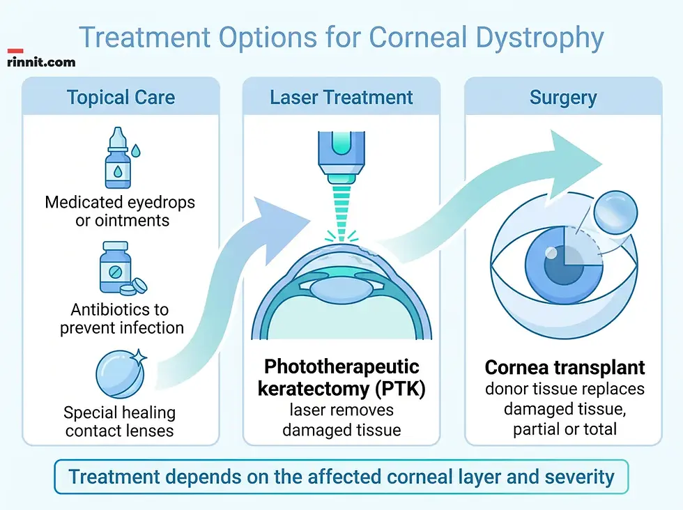
Treatment depends on the affected corneal layer and severity.
What Is the Outlook for Corneal Dystrophy?
What you can expect varies quite a bit. Most people get a diagnosis for one of these conditions by the time they reach age 20. Corneal dystrophies are usually progressive: their effects develop gradually over time, usually over years or even decades. Some people go years without knowing they have one of the conditions because they have no early symptoms, and some people never develop symptoms at all. Because experiences vary so widely, it is a good idea to ask your eye care specialist what to expect for your specific case. [1]
How Long Does Corneal Dystrophy Last?
Corneal dystrophies are lifelong conditions; you have them when you are born. Corneal transplants may "cure" these diseases, but it is not a guarantee. Many people who receive a transplant eventually develop the disease in the transplanted cornea, and further treatment, up to and including another transplant, may be necessary. Corneal dystrophies often cause at least some vision loss, but it is rare for that loss to turn into total blindness (the inability to detect light). [1]
Scenario | What to Expect |
Early years | Many people notice nothing; diagnosis often by age 20 |
Progression | Gradual, over years or decades |
With treatment | Symptoms managed, vision supported |
After cornea transplant | Lifelong immune suppression likely needed; rejection still possible |
Total blindness | Rare |
Can Corneal Dystrophy Be Prevented?
Corneal dystrophies are genetic. That means there is no way to prevent them or reduce your risk of having them. [1] What you can control is regular follow-up care, taking medications as prescribed, and getting symptoms treated early.
How Do You Take Care of Yourself With Corneal Dystrophy?
If you have corneal dystrophy, you should see your eye care specialist for regular follow-up visits as recommended. They can monitor your condition, track tissue changes and symptoms, and update your treatment plan so it stays as effective as possible for as long as possible. [1]
If your specialist prescribes medications for your eyes, take them as prescribed. Medications can limit symptoms or slow down tissue changes and damage. Taking medications exactly as prescribed is critical if you receive a corneal transplant, because it helps your body avoid rejecting the transplanted tissue, which helps you avoid severe complications and even permanent vision loss. [1]
When Should You See an Eye Care Specialist?
Seek evaluation if you notice persistent blurred or hazy vision, light sensitivity, eye pain, or the feeling that something is stuck in your eye. Anyone with a family history of corneal dystrophy should ask an eye care specialist whether monitoring makes sense, since the conditions are genetic and affect both eyes. Also talk to your specialist if treatments cause side effects or seem less effective. [1]
What questions should you ask your eye care specialist about corneal dystrophy?
Question | Why It Matters |
What type of corneal dystrophy do I have? | Determines your specific treatment path |
What treatment(s) do you recommend? | Clarifies your options based on layer and severity |
Are there any symptoms that mean I should get emergency medical care? | Helps you recognize warning signs |
What's the long-term outlook for my condition? | Sets realistic expectations |
Is there anything I can do to help improve my outlook? | Identifies your role in managing the condition |
Conclusion
Corneal dystrophy is a family of more than 20 genetic eye diseases you are born with, all of which cause tissue buildup in the cornea and make clear vision harder over time. They cannot be prevented, but they do not usually lead to total blindness, and a range of treatments, from eyedrops and special contact lenses to laser removal of damaged tissue and cornea transplants, can protect your sight for decades. If your vision has been getting hazier, your eyes water or ache after sleep, or corneal dystrophy runs in your family, book an eye exam with an ophthalmologist or optometrist so a specialist can map, measure, and test your corneas while your vision is still on your side.
To learn about the wider group of cornea problems, see our guide to corneal disease.
Frequently Asked Questions
What is corneal dystrophy in simple terms?
Corneal dystrophy is an umbrella term for over 20 genetic eye diseases present from birth that damage or disrupt repairs of the cornea, the clear front layer of the eye. They disrupt how light passes through and usually cause gradually worsening vision over years.
Is corneal dystrophy genetic?
Yes. All forms involve DNA changes. Some changes are inherited from one or both biological parents, others develop spontaneously (de novo), and a few happen for reasons experts do not fully understand yet.
What are the four types of corneal dystrophy?
The types are classified by the corneal layer affected: epithelial and subepithelial, epithelial-stromal, stromal, and endothelial dystrophies. Fuchs endothelial corneal dystrophy is the most common, accounting for about 39% of all U.S. corneal transplants.
What does corneal dystrophy feel like?
Common symptoms include blurred or hazy vision, reduced visual clarity despite glasses or contacts, a feeling that something is stuck in the eye, eye pain or irritation (often after sleep), light sensitivity, watery eyes, and rapid uncontrolled eye movements (nystagmus).
Can corneal dystrophy cause blindness?
Corneal dystrophy can cause severe vision loss that affects driving and reading, but it does not cause total blindness (the inability to detect light).
At what age does corneal dystrophy start?
Most people receive a diagnosis by age 20, and many types develop during childhood. A few forms, such as Schnyder, Pre-Descemet, epithelial basement membrane, and Fuchs dystrophies, typically appear in adulthood.
How is corneal dystrophy diagnosed?
Eye care specialists use an eye exam with a slit lamp microscope, corneal topography, pachymetry (thickness measurement), genetic testing, OCT scans, and occasionally a corneal biopsy to confirm the diagnosis and type.
How is corneal dystrophy treated?
Treatment depends on the affected layer and severity. Options include medicated eyedrops, antibiotics, special contact lenses, phototherapeutic keratectomy (laser removal of damaged tissue), and cornea transplantation from a deceased donor.
Is a cornea transplant a cure for corneal dystrophy?
Not always. Transplants may cure the disease, but many people eventually develop the dystrophy in the transplanted cornea and may need further treatment, including another transplant.
Can corneal dystrophy be prevented?
No. The conditions are genetic, so there is no way to prevent them or reduce the risk of having them. Regular eye care remains the best management strategy.
What should I ask my eye care specialist about corneal dystrophy?
Ask what type you have, what treatments are recommended, which symptoms mean you need emergency care, your long-term outlook, and what you can do to improve it.
References
Cleveland Clinic. Corneal Dystrophy. Cleveland Clinic. Medically reviewed; last updated September 24, 2024. https://my.clevelandclinic.org/health/diseases/corneal-dystrophy
Weiss JS, Møller HU, Aldave AJ, et al. IC3D classification of corneal dystrophies, edition 2. Cornea. 2015;34(2):117-159. PMID 25564336. https://doi.org/10.1097/ICO.0000000000000307
Ong Tone S, Kocaba V, Böhm M, et al. Fuchs endothelial corneal dystrophy: the vicious cycle of Fuchs pathogenesis. Progress in Retinal and Eye Research. 2021;80:100863. PMID 32438095. https://doi.org/10.1016/j.preteyeres.2020.100863
Nagpal R, Maharana PK, Roop P, et al. Phototherapeutic keratectomy. Survey of Ophthalmology. 2020;65(1):79-108. PMID 31306672. https://doi.org/10.1016/j.survophthal.2019.07.002
Medical disclaimer
This article is for general educational purposes only and is not a substitute for professional medical advice, diagnosis or treatment. Always consult a qualified eye care provider for diagnosis and treatment decisions.



Comments