Understanding Retinal Hemorrhage: Causes, Types, and Vision Health

Updated: 3 days ago
Medically reviewed by Dr. Baraa Alnahhal, MD · Last reviewed: October 2026
A retinal hemorrhage is bleeding within the retina caused by damaged blood vessels. [1] Many are found without any symptoms, but some cause sudden vision loss or blurry sight. [1] Common causes include trauma, vascular disease such as high blood pressure, and autoimmune disorders. [1] Treatment ranges from observation for small stable bleeds to injections, laser therapy or surgery for severe, vision-threatening bleeding. [1]
Quick Answer
A retinal hemorrhage is the medical term for bleeding in the retina, the light-sensitive tissue at the back of the eye. It happens when a blood vessel in the retina is damaged or breaks. [1] Some bleeds are harmless and clear on their own, while others can cause significant vision changes or permanent vision loss if the cause is not addressed. [1]
What the research says
Newborn retinal hemorrhages are common and clear quickly. A systematic review of 13 studies covering 1,777 infants found retinal hemorrhage in 25.6% of babies born by spontaneous vaginal delivery, 42.6% after vacuum extraction (odds ratio 2.75, 95% CI 1.32 to 5.70) and 52% after forceps-and-vacuum delivery; 83% had resolved within 10 days (Watts et al., Journal of AAPOS, 2013) [2].
A hemorrhage after the first month of life is not explained by birth. In 149 healthy newborns examined within 30 hours of birth, 34% had intraretinal hemorrhage (7% after cesarean section, 33% after spontaneous vaginal delivery, 75% after vacuum-assisted delivery); 86% of affected eyes had cleared by 2 weeks and none was detected at 4 weeks, so the authors advise suspecting causes other than birth when hemorrhage is found in older infants (Emerson et al., Ophthalmology, 2001) [3].
For severe bleeding into the vitreous, treatment choice is individualized. In a randomized trial of 205 adults with vitreous hemorrhage from proliferative diabetic retinopathy, initial aflibercept injections and initial vitrectomy with panretinal photocoagulation gave similar vision over 24 weeks (mean letter score 59.3 vs 63.0; adjusted difference -5.0, 95% CI -10.2 to 0.3, P = 0.06), and by 2 years both groups averaged about 20/40 vision (Antoszyk et al., JAMA, 2020) [4].
What is a retinal hemorrhage and which parts of the eye are involved?
A retinal hemorrhage occurs when blood leaks from damaged vessels into the layers of the retina. The retina converts light into signals that the brain interprets as images, so any disruption to this tissue can threaten sight. Most people have a hemorrhage in only one eye at a time (unilateral), although both eyes can be affected [1], particularly with conditions that involve the whole body.
The effect on vision depends on how much bleeding there is and where it sits. Some people never know they have a retinal hemorrhage [1] because it is found during a routine eye exam. Extensive bleeding, or bleeding that involves the center of vision, can cause lasting damage if the underlying cause is not treated.
Feature | Unilateral Hemorrhage | Bilateral Hemorrhage |
Occurrence | More common; affects one eye | Less common; affects both eyes |
Typical Cause | Trauma or eye-specific problems | Often systemic conditions such as hypertension |
Vision Impact | Affects the involved eye | Can affect vision in both eyes |
What are the types of retinal hemorrhage?
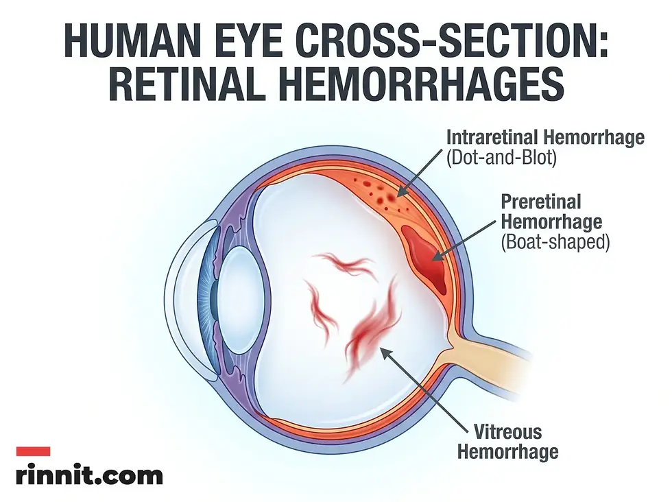
Eye care specialists classify retinal hemorrhages by the layer of the retina they involve and by their shape. [1] This helps point to the likely cause and the right treatment.
Intraretinal hemorrhages: Bleeding within the retinal tissue itself, often called "dot-and-blot" hemorrhages. [1]
Retinal nerve fiber layer hemorrhages: Bleeding in the layer of nerve cells that connects the retina to the optic nerve. Flame, disc (Drance) and splinter hemorrhages belong here. [1]
Preretinal hemorrhages: Bleeding in the membrane that separates the retina from the vitreous cavity, often boat-shaped or D-shaped. [1]
Vitreous hemorrhages: Bleeding from the retina into the vitreous, the clear gel that fills the center of the eyeball. [1]
What are the symptoms and warning signs of retinal hemorrhage?
Many retinal hemorrhages cause no symptoms. [1] When symptoms do occur, vision usually gets worse quickly enough to notice. [1] Because these changes can signal a serious underlying problem, see an eye care specialist promptly for any change in your vision.
What causes retinal hemorrhage and who is at risk?
Retinal hemorrhages are caused by injury or by health conditions that damage blood vessels over time. [1] Anyone can have one, and adults over 40 are more likely to develop age-related eye problems that cause bleeding. [1] Finding the root cause is the first step in management.
Trauma. Any injury to the head or eyes can damage retinal vessels. [1] Other traumatic triggers include shaken baby syndrome, rapid pressure changes from altitude sickness or ascending too quickly from a deep dive, and carbon monoxide poisoning. [1]
Vascular disease. Conditions that damage or narrow blood vessels are among the most common drivers in adults. These include high blood pressure, high cholesterol, atherosclerosis, retinal vein occlusion, postpartum preeclampsia and anemia. [1] Diabetes can also damage retinal vessels, and people who take blood thinners such as aspirin or warfarin have a higher risk of bleeding. [1]
Autoimmune and other disorders. Autoimmune conditions such as lupus and vasculitis can damage the retina. [1] Leukemia and infections such as bacterial endocarditis can also cause retinal hemorrhages. [1]

How is retinal hemorrhage diagnosed?
Retinal hemorrhage diagnosis starts with an eye exam, in which the specialist examines the inside of the eye and performs a visual acuity test. [1] If the cause is unclear or the damage is extensive, further tests may be used:
Blood tests: To look for signs of infection or to check cholesterol. [1]
Angiography: A specialized imaging test that maps blood flow through the retinal vessels.
MRI: Used when trauma or a condition elsewhere in the body is suspected.
How is retinal hemorrhage managed and treated?
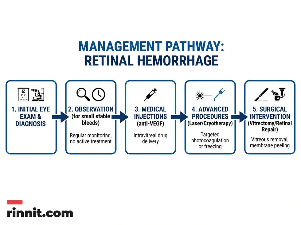
Treatment depends on the cause, how much vision is affected and how severe the bleeding is. [1] Many people need no treatment: a small hemorrhage that is not worsening and not affecting vision is usually just monitored with follow-up exams while it heals. [1]
When treatment is needed, options include:
Laser or cryotherapy: To seal leaking vessels and repair damaged retinal tissue.
Injections: Medicines such as anti-VEGF drugs or corticosteroids injected into the eye to reduce inflammation [1] and control abnormal vessels.
Surgery: In more complex cases, to reattach the retina, remove blood from the eye or normalize pressure inside the eye.
Many people make a full recovery, even if they need surgery, but permanent damage and low vision are possible. [1] Your specialist may ask you to avoid over-the-counter NSAIDs and other blood-thinning medicines while you recover. [1]
How can you protect your eyes from retinal hemorrhage?
The best way to prevent retinal hemorrhage is to prevent its causes by managing diabetes, blood pressure and cholesterol, eating healthily, not smoking and having regular eye exams. [1]
Manage diabetes, high blood pressure and high cholesterol [1], and take prescribed medicines as directed.
Eat a healthy diet low in saturated and trans fats, cholesterol, sodium and sugar, exercise regularly and keep a healthy weight. [1]
Do not smoke. [1]
Have regular eye exams: usually every 5 to 10 years under age 40, every 2 to 4 years from 40 to 54, and every 1 to 3 years from 55. People with diabetes need exams more often. [1]
Conclusion
A retinal hemorrhage is a significant finding that needs careful follow-up, even when it causes no symptoms. Managing conditions such as high blood pressure and diabetes, and seeking prompt care after an eye injury, lowers the risk of permanent vision loss. Early detection is the most effective way to protect long-term eye health.
Next step: If you notice sudden vision loss, dark spots or new flashes and floaters, seek emergency care right away. Regular eye exams help catch silent warning signs of retinal disease.
Frequently Asked Questions
Can a retinal hemorrhage heal on its own?
Yes. Many small retinal hemorrhages that are stable and do not affect vision heal without treatment. Your eye care specialist will usually arrange follow-up exams to make sure the bleeding is clearing.
Is a retinal hemorrhage a sign of a stroke?
A retinal hemorrhage is not a stroke, but it can point to vascular problems such as high blood pressure or atherosclerosis, which are stroke risk factors. A broader medical check-up is often recommended.
How long does it take for a retinal hemorrhage to clear?
It varies with the size, type and cause of the bleed. Small bleeds often clear over a few weeks, while larger vitreous hemorrhages can take longer and sometimes need surgery. Your eye specialist can give an estimate for your case.
Can exercise cause a retinal hemorrhage?
Normal exercise is not usually a concern. Intense straining can occasionally trigger bleeding in the eye, and the risk is higher with fragile blood vessels or uncontrolled high blood pressure. See an eye specialist if you notice any change in your vision.
Are babies at risk for retinal hemorrhages?
Newborn retinal hemorrhages are common, especially after assisted deliveries. In one systematic review, about 26% of babies born by spontaneous vaginal delivery and 43% of those born by vacuum extraction had them, and most cleared within 10 days. Your baby's doctor can advise if you have concerns.
What is the difference between a subconjunctival and a retinal hemorrhage?
A subconjunctival hemorrhage is a broken blood vessel on the surface of the eye and is often visible as a red patch. A retinal hemorrhage happens at the back of the eye and cannot be seen without special equipment.
Can blood thinners cause eye bleeding?
Blood thinners such as aspirin and warfarin are linked to a higher risk of retinal hemorrhage. Do not stop a prescribed blood thinner without talking to your doctor, and report any change in vision promptly.
What is a flame hemorrhage?
A flame hemorrhage is a type of bleeding in the retinal nerve fiber layer. Its feathered, flame-like shape comes from blood following the path of the nerve fibers.
When is a retinal hemorrhage an emergency?
Go to the emergency room if you suddenly lose vision in one or both eyes, have severe eye pain, or notice new flashes or floaters.
Does high cholesterol affect the eyes?
Yes. High cholesterol can contribute to atherosclerosis, which damages blood vessels throughout the body, including those in the retina, and raises the risk of retinal hemorrhage.
References
Cleveland Clinic. Retinal Hemorrhage. Cleveland Clinic. Last updated June 19, 2023. https://my.clevelandclinic.org/health/diseases/25106-retinal-hemorrhage
Watts P, Maguire S, Kwok T, et al. Newborn retinal hemorrhages: a systematic review. Journal of AAPOS. 2013;17(1):70-78. PMID 23363882. https://doi.org/10.1016/j.jaapos.2012.07.012
Emerson MV, Pieramici DJ, Stoessel KM, et al. Incidence and rate of disappearance of retinal hemorrhage in newborns. Ophthalmology. 2001;108(1):36-39. PMID 11150261. https://doi.org/10.1016/s0161-6420(00)00474-7
Antoszyk AN, Glassman AR, Beaulieu WT, et al. Effect of intravitreous aflibercept vs vitrectomy with panretinal photocoagulation on visual acuity in patients with vitreous hemorrhage from proliferative diabetic retinopathy: a randomized clinical trial. JAMA. 2020;324(23):2383-2395. PMID 33320223. https://doi.org/10.1001/jama.2020.23027
Medical disclaimer
The information provided in this article is for educational purposes only and should not be used as a substitute for professional medical advice, diagnosis, or treatment. Always seek the advice of your physician or other qualified health provider with any questions you may have regarding a medical condition.



Comments