Understanding Poikilocytosis: Abnormal Red Blood Cell Shapes

Updated: 2 days ago
Medically reviewed by Dr. Baraa Alnahhal, MD · Last reviewed: October 2026
Poikilocytosis is the medical term for abnormally shaped red blood cells, affecting more than 10% of a blood sample. Abnormal shapes can prevent red blood cells from carrying oxygen efficiently, often leading to symptoms of anemia. The condition can be inherited (like sickle cell disease) or acquired (such as nutritional deficiencies or liver disease), and it is primarily diagnosed through a peripheral blood smear, where a specialist examines cell shapes under a microscope.
Quick Answer: What is poikilocytosis?
Poikilocytosis is a condition in which abnormally shaped red blood cells, such as teardrops, sickles or spheres, are present in the bloodstream instead of round, disk-shaped cells. It often signals an underlying issue such as anemia, nutritional deficiencies or inherited genetic conditions. Misshapen cells struggle to carry oxygen, causing fatigue, weakness and other symptoms. [1]
What the research says
Reviewing red cell shape on a blood smear can be done with high agreement between methods. In a multicenter study of 882 blood smears at four sites, an automated cell-locating device with reviewer verification agreed with manual microscopy 92.1% of the time for poikilocytosis and 94.7% for anisocytosis (Jayaprakash et al., Indian J Pathol Microbiol, 2026).
Spherocytes on a smear usually need confirmatory testing. A review of laboratory diagnosis of hereditary spherocytosis notes that the traditional osmotic fragility test has low sensitivity and specificity, and that current guidelines recommend flow cytometry (the eosin-5'-maleimide binding test) as a screening test (Farias, Clin Chem Lab Med, 2017).
Some abnormal cell shapes in newborns are temporary. In a series of 9 infants with infantile pyknocytosis, a cause of transient neonatal hemolytic anemia, hemolysis resolved on its own in every case, taking a median of 33 days, although all 9 needed phototherapy and transfusions (Hu et al., Arch Pathol Lab Med, 2026).
Research summaries are based on article abstracts retrieved from PubMed.
Why do red blood cells have different shapes in poikilocytosis?
Normal red blood cells are uniform in size and shape, resembling a round disk with an indentation in the center. This specific structure is optimized for flexibility and oxygen transport. In poikilocytosis, a significant portion of these cells, typically more than 10%, exhibit structural abnormalities. [1]
Healthcare providers categorize these abnormal cells, known as poikilocytes, based on their specific visual characteristics. Identifying these shapes is a critical step in determining the underlying cause of a patient's symptoms. [1]

What are the common types of poikilocytes in poikilocytosis?
The specific shape of an abnormal red blood cell can often point toward a particular medical condition. Medical professionals use a variety of terms to describe these unique structures.
Cell Type | Visual Feature | Common Associated Conditions |
Acanthocytes | Spikes of varying lengths | Liver disease, splenectomy reaction |
Codocytes | Bullseye appearance | Thalassemia, iron deficiency, liver disease |
Dacrocytes | Teardrop shape | Myelofibrosis, megaloblastic anemia |
Drepanocytes | Crescent or sickle shape | Sickle cell disease |
Elliptocytes | Oval or cigar shape | Hereditary elliptocytosis, iron deficiency |
Spherocytes | Spherical or ball-like | Hereditary spherocytosis, autoimmune disorders |
While some shapes are highly specific, it is common for multiple abnormal cell types to be present simultaneously in conditions like thalassemia or hemolytic anemia. [1]
What are the symptoms and clinical effects of poikilocytosis?
Because poikilocytosis often leads to anemia, the symptoms are primarily related to a lack of oxygen in the body's tissues. Patients frequently experience systemic weakness and persistent fatigue that can interfere with daily activities. [1]
Commonly Reported Symptoms:
Shortness of breath (dyspnea)
Fast or irregular heartbeat (arrhythmia)
Dizziness and frequent headaches
Pale skin and occasional chest pain
A "whooshing" sound in the ears (pulsatile tinnitus) [1]
If left untreated, the chronic lack of healthy red blood cells can lead to more severe complications, including organ failure or developmental delays in children. [1]

What underlying conditions cause poikilocytosis?
Poikilocytosis is a sign of an underlying condition rather than a disease itself. These causes are generally divided into inherited and acquired categories. [1]
Inherited Conditions: These are often linked to genetic codes that affect red blood cell development. Examples include sickle cell disease, thalassemia, and hereditary spherocytosis. In these cases, cells often break down prematurely, leading to hemolytic anemia. [1]
Acquired Conditions: These develop over time and can often be reversed. Common triggers include deficiencies in iron, Vitamin B12, or folate. Other causes include chronic kidney or liver disease, certain infections, and reactions to specific medical procedures like a splenectomy. [1]
How is poikilocytosis diagnosed and tested?
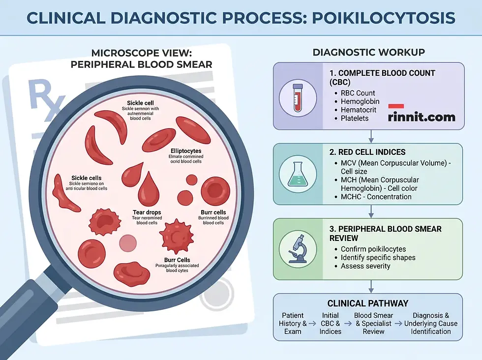
The primary diagnostic tool for poikilocytosis is the peripheral blood smear (PBS). During this test, a laboratory technologist examines a blood sample under a microscope to identify and count abnormal cell shapes. [1]
This is typically performed alongside a complete blood count (CBC), which provides data on the average size (MCV) and hemoglobin content (MCH) of the red blood cells. Providers may also order tests for vitamin levels and liver function to pinpoint the exact cause of the cell irregularities. [1]
Conclusion
Poikilocytosis serves as a vital clinical indicator of a person's hematologic health. While the presence of abnormally shaped red blood cells can be concerning, identifying the specific types of poikilocytes allows healthcare providers to diagnose and treat the root cause effectively. Whether the cause is a simple nutritional deficiency or a complex inherited trait, early detection and management are key to preventing long-term complications and restoring the body's ability to transport oxygen efficiently.
Frequently Asked Questions
What does poikilocytosis mean on a blood test?
Poikilocytosis means that more than 10% of your red blood cells have abnormal shapes, which may indicate an underlying condition like anemia.
Is poikilocytosis a serious condition?
Poikilocytosis is a sign of an underlying issue. While many causes are easily treated, untreated poikilocytosis can lead to complications like organ failure.
Can diet cause abnormally shaped red blood cells?
Yes, deficiencies in iron, Vitamin B12, and folate are common causes of acquired poikilocytosis.
What is the difference between poikilocytosis and anisocytosis?
Poikilocytosis refers to abnormal shapes, while anisocytosis refers to red blood cells of unequal sizes.
How is poikilocytosis treated?
Treatment depends entirely on the cause, ranging from dietary supplements for deficiencies to specialized management for inherited conditions.
Can children have poikilocytosis?
Yes, and if left untreated, it can slow a child's growth and development.
What are teardrop cells?
Also called dacrocytes, these are red blood cells that are pointed at one end, often seen in conditions like myelofibrosis.
Do abnormal cell shapes always mean I have anemia?
While highly common, poikilocytosis is an indicator that your cells may not be carrying oxygen efficiently, which is the hallmark of anemia.
Can a blood transfusion cause poikilocytosis?
Yes, a reaction to a blood transfusion is one of the potential acquired causes of abnormal cell shapes.
How long does it take to correct abnormal cell shapes?
The timeline depends on the cause; for example, nutritional deficiencies may resolve once levels are restored, while inherited conditions require lifelong management.
References
Cleveland Clinic. Poikilocytosis. Cleveland Clinic Health Library. Last updated March 2, 2023. https://my.clevelandclinic.org/health/diseases/24793-poikilocytosis
Jayaprakash CS, Rao S, Nair RP, et al. Clinical evaluation of AI100 with Shonit versus manual microscopy. Indian J Pathol Microbiol. 2026;69(1):87-96. PMID: 41553102. https://doi.org/10.4103/ijpm.ijpm_134_25
Farias MG. Advances in laboratory diagnosis of hereditary spherocytosis. Clin Chem Lab Med. 2017;55(7):944-948. PMID: 27837594. https://doi.org/10.1515/cclm-2016-0738
Hu AY, Coffey AM, Punia JN, et al. Infantile pyknocytosis revisited: possible familial trend in a study of 9 patients. Arch Pathol Lab Med. 2026;150(2):167-171. PMID: 40624797. https://doi.org/10.5858/arpa.2024-0504-OA
Medical disclaimer
This article is for informational purposes only and is not a substitute for professional medical advice, diagnosis, or treatment. Always seek the advice of your physician or other qualified health provider with any questions you may have regarding a medical condition.



Comments