Renal Osteodystrophy (CKD-MBD): Symptoms, Causes, Types, and Treatment

Updated: 2 days ago
Medically reviewed by Dr. Baraa Alnahhal, MD · Last reviewed: September 2026
TL;DR: Renal osteodystrophy is a bone-weakening condition caused by chronic kidney disease (CKD). It occurs when failing kidneys disrupt the balance of calcium, phosphorus, and hormones like calcitriol. Common signs include bone pain and fractures, though early stages are often asymptomatic. While a kidney transplant is the only cure, management involves dietary changes, specialized medications, and occasionally surgery to protect bone density.
Quick Answer
What is renal osteodystrophy? Renal osteodystrophy, often called CKD-MBD (Chronic Kidney Disease and Mineral Bone Disorder), is a complication where kidney dysfunction leads to mineral and hormone imbalances that damage bone tissue. Healthy kidneys regulate calcium and phosphorus levels and produce calcitriol (active vitamin D). When kidneys fail, these levels fluctuate, causing bones to either break down too quickly or fail to renew. This results in weak, brittle bones prone to pain and fractures.
Understanding Renal Osteodystrophy
Renal osteodystrophy is a complex bone disease specifically affecting individuals with chronic kidney disease (CKD). The term combines "renal" (kidney), "osteo" (bone), and "dystrophy" (damage or weakness). Healthcare providers often refer to this condition as CKD-MBD, which encompasses the bone disease along with related cardiovascular complications.
Healthy kidneys play a vital role in skeletal health by maintaining precise levels of calcium and phosphorus in the bloodstream. They also activate vitamin D into calcitriol, a hormone essential for bone maintenance. When kidney function declines, these regulatory processes fail, leading to significant bone damage.

The Three Main Types of Renal Osteodystrophy
Bone health depends on a constant cycle of "turnover," where old tissue is replaced by new tissue. Renal osteodystrophy occurs when this cycle is disrupted, moving either too fast or too slow.
Type | Bone Turnover Rate | Primary Cause | Key Characteristics |
Osteitis Fibrosa | Very High | High parathyroid hormone (PTH) | Bone tissue breaks down too fast; can form fibrous cysts. |
Osteomalacia | Low Formation | Vitamin D deficiency or metal toxicity | Bone breaks down but new tissue does not form; bones become spongy. |
Adynamic Bone Disease | Very Low | Low parathyroid hormone (PTH) | Bone tissue fails to renew itself; often linked to over-treatment. |
A fourth type, Mixed Renal Osteodystrophy, presents signs of both high-turnover osteitis fibrosa and low-formation osteomalacia simultaneously.
Symptoms and Clinical Signs
In the early stages, renal osteodystrophy is frequently asymptomatic. Patients may have significant bone damage before noticing any physical changes. As the condition progresses, the primary symptoms become more apparent and can significantly impact quality of life.
Persistent Bone Pain: A deep, aching sensation often felt in the joints or long bones.
Frequent Fractures: Broken bones occurring from minor impacts or without a clear injury.
Growth Delays: In children, the condition can lead to rickets and stunted physical development.
Skeletal Deformities: Advanced cases may result in hunched shoulders, a curved spine, or shortened fingers.

Causes and Risk Factors
The root cause of renal osteodystrophy is chronic kidney disease. The kidneys' inability to filter waste and regulate hormones creates a cascade of metabolic issues that directly attack bone integrity.
Primary Biological Triggers
Phosphorus Buildup: Failing kidneys cannot remove excess phosphorus, which pulls calcium out of the bones.
Calcitriol Deficiency: Low levels of active vitamin D prevent the body from absorbing calcium from food.
Hormonal Imbalance: The parathyroid glands may overproduce hormones to compensate for low blood calcium, further stripping minerals from the skeleton.
Who is at Risk?
Individuals with advanced CKD or those undergoing long-term dialysis are at the highest risk. Other contributing factors include diabetes, high blood pressure, heart disease, and a family history of kidney failure. Long-term use of nonsteroidal anti-inflammatory drugs (NSAIDs) can also accelerate kidney decline.
Diagnosis and Diagnostic Testing
Because the disease is often silent in its early stages, regular screening is essential for anyone diagnosed with chronic kidney disease. Providers use a combination of laboratory tests and advanced imaging to assess bone health.
Test Category | Specific Test | Purpose |
Laboratory | Blood Tests | Measures calcium, phosphorus, Vitamin D, and PTH levels. |
Imaging | DXA Scan | Measures bone density and predicts fracture risk. |
Imaging | X-ray / CT / MRI | Visualizes structural changes in the bones and joints. |
Cardiovascular | Echocardiogram | Checks for calcification in the heart and blood vessels. |
Specialized | Bone Biopsy | Analyzes bone structure directly (used in complex cases). |
Management and Treatment Pathways
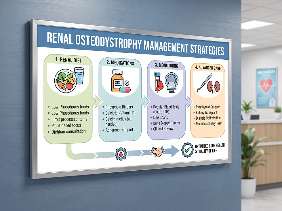
Treatment strategies focus on balancing mineral levels and slowing bone loss. While these methods cannot cure the disease, they are critical for preventing debilitating fractures and pain.
Dietary and Lifestyle Adjustments
The first line of defense is often a low-phosphorus diet. Patients are advised to limit foods high in phosphorus, such as certain dairy products, nuts, and processed meats. Strict adherence to a kidney-friendly eating plan is essential for long-term stability.
Medical Interventions
Medications are used to mimic the regulatory functions of healthy kidneys. These include phosphate binders to prevent mineral absorption in the gut and synthetic calcitriol to support calcium levels. In cases of high bone turnover, PTH-lowering drugs may be prescribed.
Surgical Options
If medication fails to control parathyroid hormone levels, a parathyroidectomy may be required. This surgery involves removing some or most of the parathyroid glands to stop the excessive loss of bone minerals.
Conclusion and Next Steps
Renal osteodystrophy is a serious but manageable complication of chronic kidney disease. By maintaining a strict treatment plan and attending regular screenings, patients can protect their skeletal health and reduce the risk of fractures. If you are living with CKD and experience new bone or joint pain, consult your healthcare provider immediately to discuss a bone health assessment.
Take Action:
Review your lab results for calcium and phosphorus levels with your specialist.
Consult a renal dietitian to optimize your low-phosphorus eating plan.
Schedule a bone density scan if recommended by your care team.
Related reading on Rinnit: Understanding Phosphate Binders: Managing Mineral Balance in Kidney Health.
For more patient information, visit the National Kidney Foundation, the American Kidney Fund, and the National Institute of Diabetes and Digestive and Kidney Diseases.
Frequently Asked Questions
Can renal osteodystrophy be reversed?
The only way to fully reverse the condition is through a successful kidney transplant, which restores normal mineral regulation.
Is renal osteodystrophy the same as osteoporosis?
No. While both cause weak bones, renal osteodystrophy is specifically caused by kidney-related metabolic imbalances, whereas osteoporosis is typically related to aging or hormonal changes like menopause.
What is the most common symptom?
Bone pain and fractures are the most common symptoms, though many people have no symptoms in the early stages.
How does phosphorus affect my bones?
High phosphorus levels in the blood signal the body to pull calcium out of your bones to balance the minerals, making the bones weak.
Why is Vitamin D important for kidney patients?
Kidneys activate Vitamin D into calcitriol. Without calcitriol, your body cannot absorb the calcium needed to maintain bone strength.
What are phosphate binders?
These are medications taken with meals to prevent your body from absorbing phosphorus from the food you eat.
Can children get renal osteodystrophy?
Yes. In children, it can cause severe growth delays and a condition known as rickets.
Is a bone biopsy always necessary?
No, bone biopsies are relatively rare and usually reserved for complex cases where blood tests and scans are inconclusive.
What foods should I avoid?
You should generally avoid high-phosphorus foods like dark sodas, processed meats, and certain dairy products, as advised by your doctor.
Can exercise help?
While exercise is generally healthy, patients with renal osteodystrophy must consult their doctor first, as their bones are more prone to breaking.
What is adynamic bone disease?
It is a type of renal osteodystrophy where bone turnover is too slow, meaning the body isn't replacing old bone tissue with new tissue.
How often should I get blood tests?
Most patients with CKD require regular blood tests, often every few months, depending on the stage of their kidney disease.
Does dialysis cure bone disease?
No, dialysis helps filter waste but does not fully replace the kidneys' hormonal functions, so bone disease can still progress.
What happens if parathyroid hormone (PTH) is too high?
High PTH levels cause the body to constantly strip calcium from the bones, leading to rapid bone loss.
Are there specific medications for bone pain?
Doctors may prescribe pain relievers, but the primary goal is to treat the underlying mineral imbalance causing the pain.
Medical Disclaimer: This information is for educational purposes only and does not constitute medical advice. Always seek the advice of your physician or other qualified health provider with any questions you may have regarding a medical condition. Never disregard professional medical advice or delay in seeking it because of something you have read here.
Comments