Left Posterior Fascicular Block (LPFB): Understanding Heart Conduction

Updated: 2 days ago
Medically reviewed by Dr. Baraa Alnahhal, MD · Last reviewed: October 2026
Left posterior fascicular block (LPFB) is a rare heart conduction disorder in which electrical signals are blocked in the left posterior fascicle. On its own it usually causes no symptoms, but it often points to underlying heart disease such as coronary artery disease, and it can be serious when combined with a block on the right side of the heart. Care focuses on treating the root cause and watching for major cardiac events [1].
Quick Answer: What is left posterior fascicular block?
Left posterior fascicular block (LPFB) is a rare conduction disorder in which the heart's electrical impulse is blocked or delayed in the left posterior fascicle, so the signal takes a different route. It usually causes no symptoms and often reflects heart disease. With a right bundle branch block, complete heart block risk is higher, and a pacemaker may be recommended. [1] [5]
What the research says
In people with bifascicular block, a few findings predict who will need a pacemaker. In a prospective study of 263 consecutive patients with chronic bifascicular block (249 analyzed, median follow-up 4.5 years), 102 needed a pacemaker, including 57 for significant atrioventricular block. Independent predictors were syncope or presyncope (hazard ratio 2.06, 95% CI 1.03–4.12), QRS width over 140 ms (HR 2.44, 95% CI 1.59–3.76), renal failure (HR 1.86, 95% CI 1.22–2.83), and an HV interval over 64 ms (HR 6.6, 95% CI 4.04–10.80); having all four was associated with a 95% probability of needing a pacemaker within 1 year. The study looked at bifascicular block as a group, not LPFB alone (Martí-Almor et al., Rev Esp Cardiol, 2010 [2]).
When fainting is unexplained, pacing guided by testing worked best. In 43 patients (mean age 78) with bifascicular block and unexplained syncope who received a pacemaker, fainting recurred in 7 (16%) over a mean follow-up of 31 months. There were no recurrences among the 17 patients paced after a positive electrophysiology study or documented paroxysmal atrioventricular block, compared with 7 of 26 (27%) who were paced empirically (P = 0.02); progression to high-degree atrioventricular block was documented in 16 patients (37%) (Kalscheur et al., Pacing Clin Electrophysiol, 2016 [3]).
What Is Left Posterior Fascicular Block?
The heart relies on a network of electrical pathways to make its chambers contract in a coordinated way. Usually, the fascicles at the front and back (posterior) of the left ventricle work together to tell it to beat. In LPFB, the posterior fascicle is blocked and cannot send electrical signals to part of the heart, so signals cannot reach the left posterior part of the lower left chamber [1].
When this happens, other signaling cells, particularly the front fascicle, step in to make the left ventricle beat. This changes where the electrical impulse goes and delays it. LPFB can be partial or complete, and it can occur on and off or without a break [1]. It is very rare in most people and more common in people with heart conditions, and isolated LPFB is an extremely rare finding even in specific patient groups [1][5].

In LPFB, the signal is blocked in the posterior part of the left ventricle and must be rerouted.
What are the common causes of left posterior fascicular block (LPFB)?
LPFB happens when there is damage to the heart, and the main causes are heart diseases and conditions. Identifying the underlying cause is the first step in effective care [1]. Coronary artery disease is the most common condition at the root of the problem, followed by high blood pressure, cardiomyopathies, and the Lev and Lenègre diseases [1][4].
Cause category | Specific heart conditions |
|---|---|
Vascular disease | Coronary artery disease (CAD): lack of blood flow to the heart because of clogged arteries, the most common underlying condition. |
Muscle disorders | Cardiomyopathy: conditions that make the heart muscle stiff, thick, or enlarged. |
Chronic conditions | Unmanaged high blood pressure, which makes it harder for the heart to pump blood and can weaken the heart muscle. |
Valve and conduction-system disease | Aortic valve disease (leaks or narrowing), Lenègre disease (inherited scarring that affects conduction), and Lev's disease (scarring of nerve fibers in the heart). |
What are the symptoms and risks of left posterior fascicular block (LPFB)?
Left posterior fascicular block (LPFB) typically causes no symptoms, though the disease causing it may produce symptoms such as chest pain or shortness of breath. People may have symptoms from the disease that is causing it, and these can signal that the underlying condition is getting worse [1]:
Chest pain: discomfort or pressure in the chest.
Shortness of breath: trouble breathing, especially with activity.
Heart palpitations: a sensation of the heart racing, fluttering, or skipping beats.
Fainting and fatigue: a sudden loss of consciousness or unusual, persistent tiredness.
LPFB becomes more significant when it occurs with a right bundle branch block, a combination known as bifascicular block. It puts you at higher risk of fatal cardiac events such as complete heart block, in which the ventricles stop receiving signals from the upper chambers. If you have a heart attack, having both blocks also increases the risk of death [1][4].
How is left posterior fascicular block diagnosed?
Healthcare providers diagnose LPFB with an electrocardiogram (ECG/EKG), which measures the heart's electrical activity. They look for very specific LPFB patterns in the results, and they will also ask about your medical history and perform a physical exam [1].
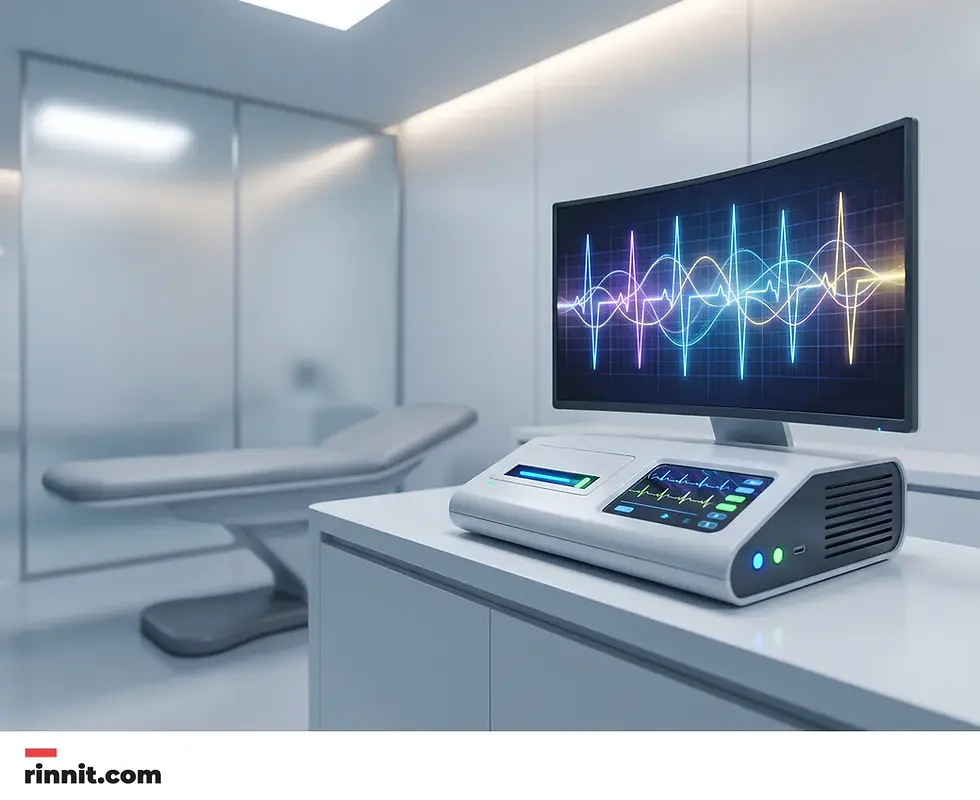
An ECG records the heart's electrical activity and is the main test used to diagnose LPFB.
A resting EKG may not show the block. Your provider may then perform an exercise stress test, which monitors the heart's activity while you are physically active and sometimes reveals blocks that did not appear at rest [1].
How is left posterior fascicular block (LPFB) managed and treated?
Left posterior fascicular block (LPFB) treatment depends on whether other heart conditions are present. If you have LPFB without other heart conditions, you will not need treatment. If it is caused by a condition such as coronary artery disease, treatment focuses on that disease [1]:
Lifestyle changes: improving diet, exercising more, and managing stress.
Medicines: drugs to control blood pressure, cholesterol, or heart rhythm.
Minimally invasive procedures or surgery: used for more advanced heart disease.
Pacemaker: if you have LPFB and a right bundle branch block along with fainting, advanced heart block or abnormal electrical testing, your provider may recommend a pacemaker, an implanted device that monitors your heart's electrical activity and sends signals to keep your heartbeat from becoming too slow [1][2][3].
To help prevent LPFB, prevent and treat the heart conditions that cause it. Routine checkups with your primary care provider can find heart problems early, and following your provider's instructions for any existing heart condition helps you avoid complications [1].
What is the bottom line on left posterior fascicular block?
Left posterior fascicular block is a rare finding that often points to broader heart health concerns. An isolated block may not be a cause for alarm, but it is an important signal for your healthcare provider to look for underlying heart disease. By managing conditions such as high blood pressure and coronary artery disease, and by following your provider's advice on monitoring and treatment, people with LPFB can maintain a good quality of life and lower the risk of serious cardiac complications.

Healthy habits and regular checkups help protect the heart.
Your next step: if you have been diagnosed with LPFB, see your primary care provider or cardiologist regularly to monitor your heart's electrical health. Ask whether you need to be concerned about LPFB, what the best treatment is for the heart condition causing it, and how often you need follow-up EKGs. See a doctor promptly if you faint or have chest pain, shortness of breath, or palpitations [1].
Frequently Asked Questions
What is LPFB?
Left posterior fascicular block (LPFB) is a rare electrical blockage in the heart's left posterior fascicle that delays signals to the left ventricle.
Is LPFB serious?
On its own, it may not be serious, but it is often a sign of underlying heart disease and can be dangerous if combined with a right bundle branch block.
What are the symptoms of LPFB?
LPFB itself usually has no symptoms. Any symptoms, such as chest pain or shortness of breath, are typically caused by the underlying heart condition.
How is LPFB diagnosed?
LPFB is primarily diagnosed using an electrocardiogram (ECG/EKG) to identify specific electrical patterns in the heart.
What causes LPFB?
The most common cause is coronary artery disease. Other causes include cardiomyopathy, high blood pressure, aortic valve disease, and certain inherited or scarring conditions.
Does LPFB require a pacemaker?
A pacemaker may be recommended if LPFB occurs alongside a right bundle branch block and there is also fainting, advanced heart block or abnormal electrical testing; the block pattern alone usually does not need one.
Can LPFB be prevented?
You can help prevent LPFB by preventing and managing the heart conditions that cause it, such as high blood pressure and coronary artery disease.
Is LPFB common?
No, LPFB is very rare in the general population but is more frequently seen in people with existing heart conditions.
What is the outlook for LPFB?
If you have no right bundle branch block or underlying heart disease, life expectancy does not change. The outlook is poor if it occurs with right bundle branch block, especially during a heart attack.
When should I see a doctor?
You should consult a healthcare provider if you have symptoms of heart disease or if LPFB is detected during a routine exam.
Related Reading
Left Anterior Fascicular Block (LAFB): Causes, Symptoms, and Heart Health covers the more common fascicular block and what it means.
Bundle Branch Block: Symptoms, Causes, and Treatment explains delays in the heart's electrical pathways and their causes.
Heart Block: Understanding Types, Symptoms, and Treatment describes heart block types and when a pacemaker is needed.
Electrocardiogram (EKG or ECG): Why It's Done, How to Prepare, What Happens and Results explains how an EKG records the heart's electrical activity.
References
Cleveland Clinic. Left Posterior Fascicular Block. Cleveland Clinic. July 22, 2025. https://my.clevelandclinic.org/health/diseases/23288-left-posterior-fascicular-block
Martí-Almor J, Cladellas M, Bazán V, et al. Novel predictors of progression of atrioventricular block in patients with chronic bifascicular block. Rev Esp Cardiol. 2010;63(4):400-408. PMID 20334805. https://pubmed.ncbi.nlm.nih.gov/20334805/
Kalscheur MM, Donateo P, Wenzke KE, et al. Long-term outcome of patients with bifascicular block and unexplained syncope following cardiac pacing. Pacing Clin Electrophysiol. 2016;39(10):1126-1131. PMID 27565449. https://doi.org/10.1111/pace.12946
Elizari MV, Acunzo RS, Ferreiro M. Hemiblocks revisited. Circulation. 2007;115(9):1154-1163. PMID 17339573. https://doi.org/10.1161/CIRCULATIONAHA.106.637389
Pérez-Riera AR, Barbosa-Barros R, Daminello-Raimundo R, et al. Left posterior fascicular block, state-of-the-art review: a 2018 update. Indian Pacing Electrophysiol J. 2018;18(6):217-230. PMID 30326266. https://doi.org/10.1016/j.ipej.2018.10.001
Medical disclaimer
This article is for informational purposes only and is not a substitute for professional medical advice, diagnosis, or treatment. The clinical information in this guide reflects source material last reviewed in July 2025.



Comments