Pulmonary Valve Stenosis: Complete Guide to Symptoms, Causes, Diagnosis, and Treatment

Updated: 2 days ago
Medically reviewed by Dr. Baraa Alnahhal, MD · Last reviewed: September 2026
TL;DR
Pulmonary valve stenosis is a narrowing of the valve between the heart's lower right chamber (the right ventricle) and the arteries that carry blood to the lungs. It usually starts as a heart defect present at birth, and in some cases develops later in life as a complication of another illness. Mild stenosis often causes no symptoms and needs only occasional checkups. Moderate or severe stenosis may require balloon valvuloplasty or valve replacement.
Quick answer: Pulmonary valve stenosis is a narrowed pulmonary valve — the one-way door between the right ventricle and the lungs — that reduces blood flow and forces the right ventricle to work harder. It is usually present at birth, is most often first detected as a whooshing heart murmur, and ranges from mild (watchful checkups) to severe (a valve-widening or replacement procedure). See a doctor if you or your child has shortness of breath, chest pain, or fainting, and call 911 or your local emergency number for sudden or unexplained chest pain.
What Is Pulmonary Valve Stenosis?
Pulmonary valve stenosis is a type of heart valve disease. It is a narrowing of the valve between the heart's lower right chamber, called the right ventricle, and the arteries that carry blood to the lungs.

Side-by-side heart diagrams comparing a normal pulmonary valve with pulmonary valve stenosis, where thick, stiff, or fused flaps leave only a small opening.
To understand the condition, it helps to know how the valve normally works. The pulmonary valve is made of three thin pieces of tissue called flaps, or cusps. These cusps open and close with each heartbeat, making sure blood moves in the right direction — from the right ventricle into the pulmonary artery and on to the lungs.
In pulmonary valve stenosis, one or more of the cusps may be stiff or thick. Sometimes the cusps are joined together, or fused. Either way, the valve cannot open fully. The smaller opening makes it harder for blood to leave the right ventricle.
Answer nugget: In pulmonary valve stenosis, one or more valve flaps (cusps) are stiff, thick, or fused, so the valve cannot open fully and blood flow to the lungs is reduced.
Pressure then increases inside the chamber. This extra strain forces the right ventricle to pump harder, and over time the muscular wall of the right ventricle becomes thicker.
The condition ranges from mild to severe. People with mild stenosis may have no symptoms at all and need only occasional health checkups. People with moderate or severe stenosis may need a procedure to repair or replace the valve.
What Causes Pulmonary Valve Stenosis?
Pulmonary valve stenosis usually results from a heart problem that is present at birth — a congenital heart defect. The pulmonary valve does not develop properly as the baby grows in the womb, and the exact cause is unclear.
In adults, pulmonary valve stenosis may also develop as a complication of another illness. Conditions that can damage the valve over time are covered in the risk factors section below.
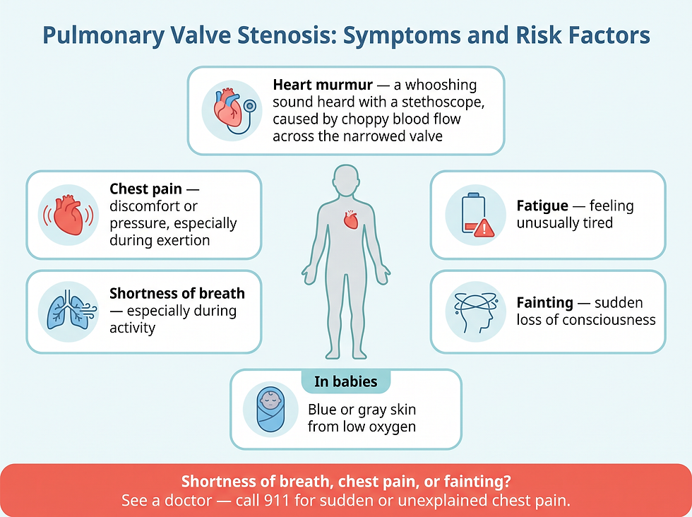
The main symptoms of pulmonary valve stenosis — heart murmur, chest pain, shortness of breath, fatigue, fainting — plus blue or gray skin in babies from low oxygen.
Symptoms of Pulmonary Valve Stenosis
Symptoms depend on how much blood flow is blocked. Many people with mild pulmonary stenosis have no symptoms. Those with more severe stenosis may first notice symptoms while exercising.
Symptoms may include the following:
Heart murmur: A whooshing sound heard with a stethoscope, caused by choppy blood flow across the narrowed valve
Fatigue: Feeling unusually tired or run down
Shortness of breath: Breathing difficulty, especially during activity
Chest pain: Discomfort or pressure in the chest
Fainting: Sudden loss of consciousness
Babies with pulmonary valve stenosis may have blue or gray skin due to low oxygen levels.
Answer nugget: Mild pulmonary valve stenosis often causes no symptoms; the most common first sign is a whooshing heart murmur heard through a stethoscope, caused by choppy blood flow across the narrowed valve.
When to See a Doctor
The guidance from the clinical literature is simple: talk to a health care provider if you or your child has shortness of breath, chest pain, or fainting.
Prompt diagnosis and treatment of pulmonary valve stenosis can help reduce the risk of complications.
Emergency note: Call 911 or your local emergency number if chest pain is sudden or cannot be explained.
Risk Factors
Four factors are associated with an increased risk of pulmonary valve stenosis.
Rubella (German measles) during pregnancy: Having rubella while pregnant increases the risk of pulmonary valve stenosis in the baby
Noonan syndrome: A condition caused by altered DNA that can lead to many problems with the heart's structure and function
Rheumatic fever: A complication of strep throat that can cause permanent damage to the heart and heart valves, raising the risk of pulmonary valve stenosis later in life
Carcinoid syndrome: A rare cancerous tumor releases chemicals into the bloodstream; some people develop carcinoid heart disease, which damages heart valves
Answer nugget: Risk factors for pulmonary valve stenosis include rubella (German measles) during pregnancy, Noonan syndrome, rheumatic fever after strep throat, and carcinoid syndrome.
Complications of Pulmonary Valve Stenosis
Left untreated, moderate and severe stenosis can lead to serious complications. Understanding these is part of why clinicians recommend timely evaluation and treatment.
Infective endocarditis: People with heart valve problems have an increased risk of bacterial infections that affect the inner lining of the heart
Arrhythmias: Irregular heartbeats are more common with pulmonary stenosis; unless the stenosis is severe, these are usually not life-threatening
Right ventricular hypertrophy: In severe stenosis, the right ventricle must pump harder, and its muscular wall thickens
Heart failure: If the right ventricle cannot pump properly, heart failure eventually develops, with symptoms such as fatigue, shortness of breath, and swelling of the legs and belly area
Pregnancy complications: Labor and delivery risks are higher for people with severe pulmonary valve stenosis than for those without it
Answer nugget: Possible complications of pulmonary valve stenosis include infective endocarditis, arrhythmias, thickening of the right ventricle (right ventricular hypertrophy), heart failure, and increased risks during labor and delivery.
How Pulmonary Valve Stenosis Is Diagnosed
Pulmonary valve stenosis is often diagnosed in childhood, but it may not be detected until later in life. A health care provider uses a stethoscope to listen to the heart, and the whooshing murmur — caused by choppy blood flow across the narrowed valve — may be the first clue.
Several tests are used to confirm the diagnosis and measure severity.
Electrocardiogram (ECG or EKG): A quick, painless test with electrodes on the chest, arms, and legs that records the heart's electrical signals; can show how the heart is beating and may reveal signs of heart muscle thickening
Echocardiogram: Uses sound waves to produce images of the heart; shows how the heart beats and pumps blood, the shape of the pulmonary valve, and how much of the valve is narrowed
Cardiac catheterization: A thin tube (catheter) is inserted into the groin and threaded through the blood vessels to the heart; dye makes the vessels visible on X-rays, and pressures within the heart are measured; severity is determined by the pressure difference between the right ventricle and the lung artery
MRI and CT scans: Sometimes used to confirm the diagnosis
Answer nugget: Pulmonary valve stenosis is usually diagnosed with an echocardiogram, which shows how much the valve is narrowed; cardiac catheterization measures the pressure difference between the right ventricle and the lung artery to grade severity.
Treatment Options
Treatment depends on how severe the stenosis is and on the person's overall health and valve anatomy.
People with mild pulmonary valve stenosis without symptoms may only need occasional health checkups. Those with moderate or severe stenosis may need a heart procedure or heart surgery.
Balloon Valvuloplasty
During balloon valvuloplasty, a provider inserts a flexible tube with a balloon on the tip into an artery, usually in the groin. X-rays help guide the catheter to the narrowed valve in the heart. The balloon inflates, making the valve opening larger, then deflates, and the catheter and balloon are removed.
Valvuloplasty may improve blood flow through the heart and reduce symptoms. However, the valve may narrow again, and some people need valve repair or replacement in the future.
Pulmonary Valve Replacement
If balloon valvuloplasty is not an option, the pulmonary valve may be replaced through open-heart surgery or a catheter procedure. If other heart problems are present, the surgeon may repair them during the same surgery.
An important lifelong detail: people who have had pulmonary valve replacement need to take antibiotics before certain dental procedures or surgeries to prevent endocarditis, the bacterial infection of the heart's inner lining.
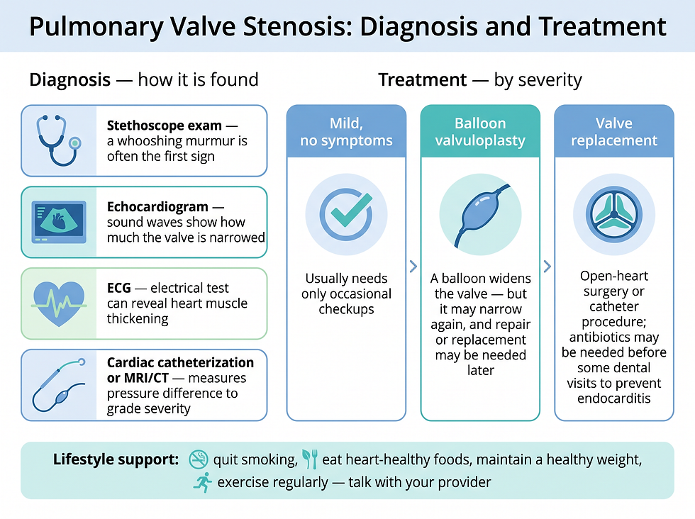
Diagnosis steps (stethoscope exam, echocardiogram, ECG, cardiac catheterization or MRI/CT) and treatment by severity: occasional checkups, balloon valvuloplasty, or valve replacement.
Answer nugget: Treatment for moderate or severe pulmonary valve stenosis is balloon valvuloplasty (widening the valve with an inflated balloon) or pulmonary valve replacement; mild cases without symptoms usually need only occasional checkups.
Self-Care and Heart-Healthy Habits
If you have valve disease, it is important to take steps to keep your heart healthy. Certain lifestyle changes can decrease the risk of developing other types of heart disease or having a heart attack. Discuss the following with your health care provider:
Quitting smoking: Removes a major strain on the heart and blood vessels
Eating a heart-healthy diet: Fruits and vegetables, low-fat dairy products, whole grains, and lean meat support heart health
Maintaining a healthy weight: Reduces extra workload on the heart
Getting regular exercise: Keeps the heart muscle and circulation strong
Preparing for Your Appointment
If you or your child has valve disease, you will likely be referred to a cardiologist — a doctor trained in evaluating and treating heart conditions.
To prepare, write down your or your child's symptoms, key personal information such as major stresses or recent illnesses, and a complete list of all medications, vitamins, and supplements. Bringing a list of questions makes the most of your time.
Useful questions to ask include: What is the most likely cause of the symptoms? What tests are needed, and do they require special preparation? Is the stenosis temporary or long lasting? What treatments are available and which is recommended? What are the risks of balloon valvuloplasty or open-heart surgery? Are there activity restrictions?
Expect your provider to ask when symptoms began, whether they come and go, whether they worsen during activity or when lying down, and whether anything seems to improve them.
Answer nugget: People with pulmonary valve stenosis are usually referred to a cardiologist; come prepared with a symptom journal, a medication list, and questions about tests, treatment options, and activity restrictions.
Conclusion and Next Steps
Pulmonary valve stenosis is a narrowing of the one-way valve between the right ventricle and the lungs — most often present at birth, first noticed as a whooshing murmur, and ranging from mild enough for watchful checkups to severe enough for balloon valvuloplasty or valve replacement.
The trigger to act: any episode of shortness of breath, chest pain, or fainting in you or your child is a signal to see a doctor, because prompt diagnosis and treatment can help reduce the risk of complications such as heart failure, arrhythmias, and endocarditis.
Before your appointment, bring a written symptom journal, a full medication list, and the questions listed above so you and your cardiologist can decide whether monitoring or a valve procedure is the right path.
Frequently Asked Questions
1. What is pulmonary valve stenosis?
Pulmonary valve stenosis is a narrowing of the pulmonary valve — the valve between the heart's lower right chamber (right ventricle) and the arteries to the lungs. Stiff, thick, or fused valve flaps reduce blood flow and make the right ventricle work harder.
2. What causes pulmonary valve stenosis?
It usually results from a heart defect present at birth: the pulmonary valve does not develop properly as the baby grows in the womb, and the exact cause is unclear. In adults, it can also develop as a complication of another illness, such as rheumatic fever or carcinoid syndrome.
3. What are the symptoms of pulmonary valve stenosis?
Mild cases may have no symptoms. Possible symptoms include a whooshing heart murmur, fatigue, shortness of breath especially during activity, chest pain, and fainting. Babies may have blue or gray skin from low oxygen.
4. How is pulmonary valve stenosis diagnosed?
A stethoscope exam often detects a whooshing murmur first. An echocardiogram shows the valve's shape and how much it is narrowed. Cardiac catheterization measures the pressure difference between the right ventricle and the lung artery to grade severity, and MRI or CT scans are sometimes used to confirm the diagnosis.
5. How is pulmonary valve stenosis treated?
Mild stenosis without symptoms usually needs only occasional checkups. Moderate or severe stenosis is treated with balloon valvuloplasty (widening the valve with an inflated balloon) or pulmonary valve replacement through open-heart surgery or a catheter procedure.
6. Can pulmonary valve stenosis come back after balloon valvuloplasty?
Yes. The valve may narrow again after valvuloplasty, and some people need valve repair or replacement later on.
7. What complications can pulmonary valve stenosis cause?
Complications include infective endocarditis, arrhythmias (usually not life-threatening unless the stenosis is severe), thickening of the right ventricle wall (right ventricular hypertrophy), heart failure, and increased risks of complications during labor and delivery.
8. Do I need antibiotics after pulmonary valve replacement?
Yes, for certain procedures. People who have had pulmonary valve replacement need to take antibiotics before some dental procedures or surgeries to prevent endocarditis.
Related Resources on Rinnit
Two related, in-depth guides may help. Read more about congenital heart defects in a parent's complete guide to pulmonary atresia with VSD, or explore our complete overview of pulmonary valve disease, covering all three valve conditions, the four disease stages, and treatment options.
Trusted External Sources
Medical Disclaimer
This article is for general educational purposes only and is not a substitute for professional medical advice, diagnosis, or treatment. Always consult a qualified health care provider about your specific condition. Call 911 or your local emergency number for sudden or unexplained chest pain, fainting, or any medical emergency.
Comments