Pulmonary Valve Disease: Complete Guide to Symptoms, Causes, and Treatment

Updated: 2 days ago
Medically reviewed by Dr. Baraa Alnahhal, MD · Last reviewed: September 2026
TL;DR
Pulmonary valve disease affects the valve between the heart's right ventricle and the pulmonary artery, the vessel that carries blood to the lungs. There are three main types — stenosis (a narrowed valve), regurgitation (a leaky valve), and atresia (a valve never formed at birth). It is usually caused by a congenital heart defect, though other illnesses can damage the valve later in life. Diagnosis often begins with a stethoscope finding of a heart murmur, and treatment ranges from simple monitoring to balloon valvuloplasty, valve repair, or valve replacement.
Quick answer: Pulmonary valve disease is any condition in which the pulmonary valve — the one-way door between the right ventricle and the lungs — fails to work properly. See a doctor if you or your child has shortness of breath, chest pain, or fainting. Call 911 or your local emergency number if chest pain is sudden or cannot be explained.
What Is Pulmonary Valve Disease?
Pulmonary valve disease is a type of heart valve disease. It affects the valve between the heart's lower right chamber and the artery that delivers blood to the lungs. That artery is called the pulmonary artery, and the valve itself is called the pulmonary valve.
A diseased or damaged pulmonary valve doesn't work the way it should. It changes how blood flows from the heart to the lungs.
To understand how the disease happens, it helps to know how the valve normally works. The pulmonary valve is like a one-way door from the lower right heart chamber, called the right ventricle, to the lungs. Blood flows through the valve into the lungs, picks up oxygen, and carries it to the body.
When the valve doesn't work as it should, the heart must work harder to pump blood to the lungs. That extra work can cause the right ventricle to become thick — a change doctors watch closely because it strains the heart over time.
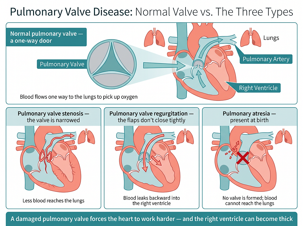
Figure 1: Side-by-side comparison of a normal pulmonary valve with the three types of pulmonary valve disease: stenosis, regurgitation, and atresia.
The Three Types of Pulmonary Valve Disease
The condition is not one single problem. There are three distinct types, and each changes blood flow in a different way.
Pulmonary valve stenosis — the valve is narrowed, which reduces blood flow from the heart to the pulmonary artery and lungs
Pulmonary valve regurgitation — the flaps of the valve don't close tightly, so blood moves backward into the right ventricle
Pulmonary atresia — present at birth (congenital heart defect); the pulmonary valve isn't formed, so a solid sheet of tissue blocks blood flow and blood can't reach the lungs to pick up oxygen
The key distinction matters for diagnosis and treatment: stenosis and regurgitation involve a valve that formed but doesn't work right, while atresia means the valve never formed at all. Pulmonary atresia is therefore classified as a congenital heart defect rather than a disease that develops later.
Symptoms of Pulmonary Valve Disease
Symptoms depend on the specific valve condition and how much blood flow is changed or blocked. Some babies are born with pulmonary valve disease, and symptoms may be noticed soon after birth. But sometimes symptoms aren't seen until later in life.

Figure 2: The main symptoms of pulmonary valve disease — chest pain, shortness of breath, fatigue, and fainting — plus blue or gray skin in babies from low oxygen.
What symptoms look like
Chest pain: Discomfort or pressure in the chest area
Shortness of breath: At rest, when active, or lying down
Fatigue: Unusual tiredness or low energy
Fainting: Loss of consciousness or feeling faint
Blue or gray skin (babies): Caused by low oxygen levels; may be harder or easier to see depending on skin color
When to see a doctor
Make an appointment for a health checkup if you or your child has shortness of breath, chest pain, or fainting. Call 911 or your local emergency number if chest pain is sudden or can't be explained. Quick diagnosis and treatment of pulmonary valve disease can help reduce the risk of complications.
Causes and Risk Factors
Usually, pulmonary valve disease is caused by a heart condition that develops before birth, called a congenital heart defect. But pulmonary valve disease may also happen later in life as a complication of another illness.
What raises the risk of pulmonary valve disease:
Congenital heart defect: Some birth defects affect the shape of the pulmonary valve and how it works
Carcinoid syndrome: A rare cancerous tumor sends chemicals into the bloodstream; can cause carcinoid heart disease, which commonly damages the pulmonary and tricuspid valves
Chest injury: An injury such as from a car accident may damage the valve and lead to regurgitation
German measles (rubella) during pregnancy: Increases the risk of pulmonary valve stenosis in the baby
Endocarditis: Infection of the lining of the heart and heart valves can damage the pulmonary valve; IV drug misuse raises the risk of endocarditis
Noonan syndrome: A genetic condition; pulmonary valve stenosis is a common heart complication
Two of these risk factors are worth highlighting. Rubella during pregnancy raises the baby's risk of pulmonary valve stenosis — one reason vaccination before pregnancy matters. And endocarditis, an infection of the heart's lining and valves, can directly damage the pulmonary valve; IV drug misuse makes endocarditis more likely.
How Pulmonary Valve Disease Is Diagnosed
Some types of pulmonary valve stenosis are diagnosed at birth or soon after. But sometimes pulmonary valve disease isn't noticed until later in life.
Diagnosis usually begins in the simplest way: a healthcare professional uses a stethoscope to listen to the heart. A whooshing sound, called a heart murmur, may be heard — often the first clue that a valve isn't working normally.
Electrocardiogram (ECG or EKG): Quick and painless test that shows how the heart is beating; electrodes attach to the chest and sometimes the arms and legs
Echocardiogram: Sound waves create images of the heart; shows blood flow through the heart and valves and can show the shape of the pulmonary valve
Chest X-ray: Shows the heart and lungs; reveals whether the heart is larger than usual or whether fluid surrounds the lungs
Cardiac catheterization: Not often used to diagnose valve disease, but may measure how severe it is; a catheter is guided to the heart and dye highlights the arteries on X-ray
MRI or CT scan: Imaging tests sometimes used to confirm pulmonary valve disease, such as stenosis
An important diagnostic nuance: cardiac catheterization is not often used to diagnose heart valve disease — its role is mainly to assess severity after other tests point to a problem.
The Four Stages of Heart Valve Disease
After testing confirms a diagnosis, the healthcare team may assign a stage. Staging helps determine the most appropriate treatment and depends on symptoms, disease severity, the structure of the valve, and blood flow through the heart and lungs.
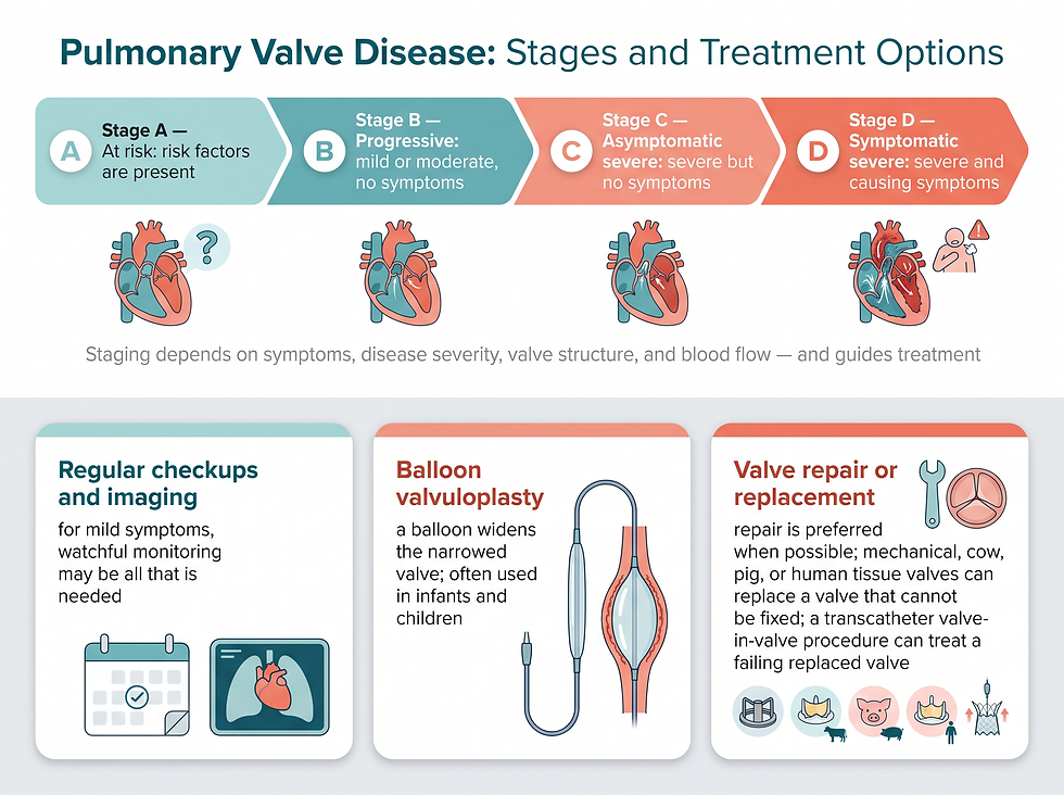
Figure 3: The four stages of heart valve disease from Stage A (at risk) through Stage D (symptomatic severe), with treatment options including monitoring, balloon valvuloplasty, and valve repair or replacement.
Stage A — At risk: Risk factors for heart valve disease are present
Stage B — Progressive: Valve disease is mild or moderate; no symptoms
Stage C — Asymptomatic severe: No symptoms, but the valve disease is severe
Stage D — Symptomatic severe: Valve disease is severe and causing symptoms
This staging system carries an important warning: Stage C disease is severe even though the person feels nothing. That is why monitoring matters — serious valve disease can be progressing silently.
Treatment Options for Pulmonary Valve Disease
Treatment depends on the symptoms, the stage of the disease, and whether the disease is getting worse. Options include regular health checkups and surgery to repair or replace the valve.
If symptoms are mild, the only treatment may be regular health checkups and imaging tests to see how the heart is working.
What determines repair versus replacement
The decision to repair or replace a damaged pulmonary valve depends on the stage of disease, symptoms, age and overall health, whether the condition is worsening, and whether surgery is needed to correct another valve or heart condition. Pulmonary valve surgery may be done at the same time as other heart valve surgeries.
Surgeons recommend pulmonary valve repair when possible, because it saves the heart valve and improves heart function.
The three main procedures
Balloon valvuloplasty: A catheter is guided to the heart, usually through the groin; a balloon on its tip is inflated to widen the valve opening, then deflated and removed. Often used for infants and children with a narrowed pulmonary valve.
Pulmonary valve replacement surgery: The valve is replaced with a mechanical valve or one made from cow, pig, or human heart tissue (a biological tissue valve). Used when the valve can't be fixed.
Transcatheter pulmonary valve replacement: A minimally invasive procedure using thin, flexible tubes and smaller surgical cuts; can also place a new valve inside a previously replaced valve that is now failing — a valve-in-valve procedure. Suits patients who qualify for minimally invasive replacement, including some with a failing prior valve replacement.
Three treatment facts stand out for people researching their options. First, repair is preferred over replacement when possible. Second, balloon valvuloplasty is the procedure most often used in infants and children with a narrowed valve — and it avoids open surgery entirely. Third, a valve-in-valve transcatheter procedure means a failing replaced valve can sometimes be treated without redoing open surgery.
Living With Pulmonary Valve Disease
For people with mild disease, active monitoring is itself a treatment plan. Regular checkups and imaging tests track how the heart is working and whether the disease is progressing through the four stages.
Anyone diagnosed with pulmonary valve disease should keep every scheduled appointment and report new symptoms — especially shortness of breath, chest pain, or fainting — without waiting for the next checkup.
Questions to ask your doctor
Which type of pulmonary valve disease do I (or my child) have, and what stage is it?
Is the valve a candidate for repair rather than replacement?
If a replacement is needed, which valve type — mechanical or biological — fits my situation?
Could balloon valvuloplasty or a transcatheter procedure be an option?
How often should imaging tests be repeated?
Conclusion: When a Heart Murmur Needs a Follow-Up
Pulmonary valve disease spans a wide range — from a narrowed valve in a child, to a leaky valve discovered in adulthood, to a valve that never formed at birth. The common thread is simple: the heart must work harder, and over time the right ventricle can thicken.
The good news is that diagnosis often starts with a routine stethoscope exam, and treatment today includes minimally invasive options that may not have existed a generation ago. If you or your child has shortness of breath, chest pain, or fainting, make an appointment for a health checkup. If chest pain is sudden or unexplained, call 911 or your local emergency number right away.
Next step: Bring this guide's question list to your appointment, and ask specifically whether repair is possible before replacement is discussed — because saving the valve, when possible, improves heart function.
Related reading: Pulmonary atresia is one type of pulmonary valve disease and is itself a congenital heart defect — learn more about how it is diagnosed and treated.
Frequently Asked Questions (FAQ)
1. What is pulmonary valve disease?
Pulmonary valve disease is a type of heart valve disease affecting the valve between the heart's lower right chamber (right ventricle) and the pulmonary artery, which delivers blood to the lungs. When the valve is damaged, it changes how blood flows from the heart to the lungs.
2. What are the three types of pulmonary valve disease?
The three types are pulmonary valve stenosis (the valve is narrowed, reducing blood flow to the lungs), pulmonary valve regurgitation (the valve flaps don't close tightly, so blood leaks backward into the right ventricle), and pulmonary atresia (the valve isn't formed at birth, blocking blood flow to the lungs).
3. What causes pulmonary valve disease?
Usually, it is caused by a congenital heart defect that develops before birth. But it can also happen later in life as a complication of another illness, such as endocarditis, carcinoid syndrome, or chest injury.
4. What are the symptoms of pulmonary valve disease?
General symptoms include chest pain, shortness of breath at rest or during activity or lying down, fatigue, and fainting. Babies born with the condition may have blue or gray skin due to low oxygen levels, which may be harder or easier to see depending on skin color.
5. How is pulmonary valve disease diagnosed?
Diagnosis typically starts with a stethoscope exam, where a whooshing sound called a heart murmur may be heard. Tests such as an ECG, echocardiogram, chest X-ray, and sometimes cardiac catheterization, MRI, or CT scans confirm the diagnosis and measure severity.
6. Is pulmonary valve disease serious?
It can be. Untreated disease forces the heart to work harder and can cause the right ventricle to become thick. Severe disease is staged from A (at risk) through D (symptomatic severe). Quick diagnosis and treatment can help reduce the risk of complications.
7. What is the treatment for pulmonary valve disease?
Treatment depends on symptoms, disease stage, and whether the disease is worsening. Mild cases may need only regular checkups and imaging. Procedural options include balloon valvuloplasty (often for infants and children), valve repair (preferred when possible), valve replacement with mechanical or biological tissue valves, and transcatheter valve-in-valve procedures.
8. Can the pulmonary valve be repaired instead of replaced?
In general, surgeons recommend pulmonary valve repair when possible, because it saves the heart valve and improves heart function. Replacement with a mechanical, cow, pig, or human tissue valve is considered when the valve can't be fixed.
References
External resources: American Heart Association — Heart Valve Disease · National Heart, Lung, and Blood Institute — Heart Valve Diseases · American College of Cardiology — Valvular Heart Disease Guidelines
Medical disclaimer: This article is for general informational purposes only and does not constitute medical advice. Always consult a qualified healthcare professional for diagnosis and treatment decisions. If you experience sudden or unexplained chest pain, call 911 or your local emergency number immediately.
Comments