Orbital Cellulitis: Recognizing the Signs of a Serious Eye Infection

Updated: 2 days ago
Medically reviewed by Dr. Baraa Alnahhal, MD · Last reviewed: September 2026
TL;DR: Orbital cellulitis is a severe bacterial infection of the soft tissues—including fat and muscles—within the eye socket (orbit). Most commonly affecting children under seven, it often originates from a sinus infection that spreads through the thin membranes separating the sinuses from the eye. Unlike superficial eyelid infections, orbital cellulitis is a medical emergency that can lead to permanent vision loss, meningitis, or brain abscesses if not treated immediately. Symptoms include painful eye movement, a bulging eye (proptosis), high fever, and significant swelling. Hospitalization for intravenous (IV) antibiotics is the standard of care, and early intervention typically results in a full recovery without long-term damage.
Life-Threatening Medical Emergency
Orbital cellulitis is a rapidly progressing infection that can threaten both vision and life. Seek immediate emergency medical care if you or your child experience:
A bulging eye (proptosis)
Pain or extreme difficulty when moving the eye
High fever accompanied by severe eyelid swelling
Sudden vision loss or significant blurring
Swelling that extends beyond the eyelid to the eyebrow or cheek
Quick answer: Orbital cellulitis is a bacterial infection of the tissues inside the eye socket, including the skin, fat, and muscles surrounding the eye. It is most prevalent in young children and is significantly more dangerous than common eyelid infections. The bacteria usually travel from the ethmoid sinuses into the orbit, causing inflammation that can compress the optic nerve and restrict blood flow. Because of the proximity to the brain, the infection carries a risk of spreading to the central nervous system. Immediate treatment with IV antibiotics in a hospital setting is required to stop the infection and prevent complications like blindness or meningitis.
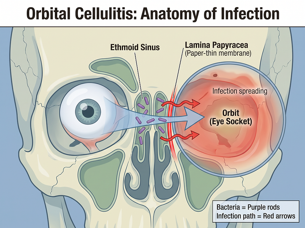
The Anatomy of Infection
The eye socket is separated from the sinuses by a paper-thin membrane, which serves as a common pathway for bacterial spread.
Ethmoid Sinus: Located beside the nose, closest to the eye. It is the primary source of most orbital infections.
Orbit (Eye Socket): The bony cavity containing the eye. It is the site of bacterial colonization and tissue swelling.
Optic Nerve: Transmits visual information to the brain. It can be damaged by pressure, leading to blindness.
Eye Muscles: Control the movement of the eyeball. They can become paralyzed, causing double vision.
Symptoms and Warning Signs
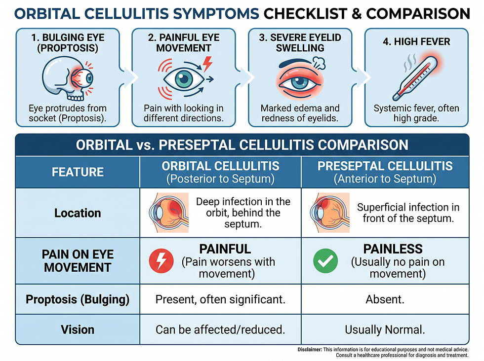
Orbital cellulitis presents with systemic and localized symptoms that distinguish it from less severe conditions.
Primary Symptoms
Bulging Eye (Proptosis): The eye appears pushed forward due to swelling behind it.
Painful Movement: Moving the eye in any direction causes significant discomfort.
Systemic Illness: High fever and a general feeling of being unwell (malaise).
Vision Changes: Blurred vision or a complete loss of sight in the affected eye.
Comparison: Orbital vs. Preseptal Cellulitis
It is critical to distinguish between these two types of cellulitis, as their risks differ significantly.
Location: Preseptal (Periorbital): Eyelid skin only; Orbital Cellulitis: Deep tissues inside the socket
Eye Movement: Preseptal (Periorbital): Normal and painless; Orbital Cellulitis: Painful or restricted
Eye Position: Preseptal (Periorbital): Normal; Orbital Cellulitis: Bulging (Proptosis)
Vision: Preseptal (Periorbital): Unaffected; Orbital Cellulitis: May be impaired
Severity: Preseptal (Periorbital): Moderate; Orbital Cellulitis: Emergency
Causes and Risk Factors
Bacteria are the sole cause of orbital cellulitis, often entering the orbit from neighboring structures.
Sinusitis: The most common cause, specifically infections of the ethmoid sinuses.
Pathogens: Staphylococcus (Staph) and Pneumococcus are frequent culprits.
Secondary Sources: Infections from the teeth, ears, or facial skin (necrotizing fasciitis).
Trauma: Eye injuries or surgical procedures that break the protective barriers of the orbit.
Age: Children under the age of 7 are at the highest risk due to their developing sinus anatomy.
Complications of Delayed Treatment
If not addressed within hours, orbital cellulitis can lead to irreversible damage.
Vision Loss: Permanent blindness caused by optic nerve compression.
Muscle Paralysis: Inability to move the eye, resulting in permanent double vision.
Intracranial Spread: The infection can move to the brain, causing meningitis or blood clots (cavernous sinus thrombosis).
Sepsis: A life-threatening systemic response to the infection spreading through the bloodstream.
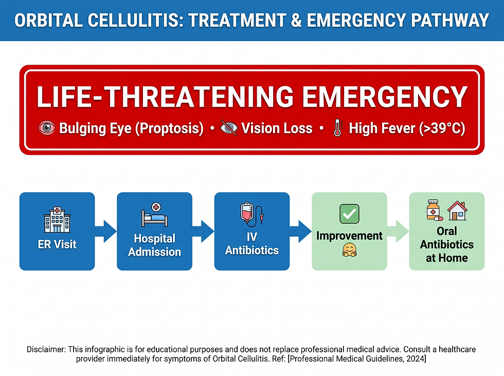
Treatment and Recovery Pathway
Because of the high risk, treatment is aggressive and typically requires a hospital stay.
Hospital Management
Patients are admitted for intravenous (IV) antibiotics, which deliver medication directly into the bloodstream for maximum speed and efficacy. A bacterial culture may be taken to ensure the chosen antibiotic is effective against the specific strain.
Surgical Intervention
While rare, surgery may be necessary to:
Drain a localized abscess (collection of pus).
Relieve dangerous pressure on the optic nerve.
Remove dead or infected tissue (debridement).
Home Care
Once symptoms improve, patients transition to oral antibiotics for several weeks. It is mandatory to complete the entire course of medication to prevent the infection from returning.
Outlook and Prognosis
With prompt medical attention, the prognosis for orbital cellulitis is excellent. Most patients, especially children, make a full recovery with no lasting impact on their vision. The key to this positive outcome is the speed of the initial ER visit and strict adherence to the antibiotic regimen.
Frequently Asked Questions (FAQ)
Is orbital cellulitis contagious?
The infection itself is not contagious like a cold, but the bacteria causing it can be spread through direct contact. However, orbital cellulitis usually develops from a person's own sinus infection.
Can orbital cellulitis cause blindness?
Yes. If the swelling is severe enough to compress the optic nerve or cut off blood flow to the eye, permanent blindness can occur.
Why is it more common in children?
Children under 7 have developing sinuses and thinner membranes between the sinuses and the eye socket, making it easier for bacteria to spread.
How long is the hospital stay for orbital cellulitis?
Most patients stay in the hospital for a few days to receive IV antibiotics until their symptoms begin to show significant improvement.
Can I treat orbital cellulitis at home?
No. Orbital cellulitis is a medical emergency that requires IV antibiotics in a hospital. Home treatment with oral pills is only appropriate after the initial infection is under control.
What is the difference between a stye and orbital cellulitis?
A stye is a small, localized bump on the eyelid. Orbital cellulitis involves deep swelling of the entire eye socket, fever, and painful eye movement.
Can a sinus infection lead to orbital cellulitis?
Yes. Sinusitis, particularly of the ethmoid sinuses, is the most common cause of orbital cellulitis.
What does a "bulging eye" look like?
The eyeball may appear to be pushed forward or look larger than the other eye because of the pressure from swelling behind it.
Is surgery always required?
No. Most cases are successfully treated with antibiotics alone. Surgery is reserved for complications like abscesses or extreme pressure.
Can orbital cellulitis affect the brain?
Yes. The infection can spread through blood vessels or membranes to the brain, potentially causing meningitis or life-threatening blood clots.
How do doctors know if it's orbital cellulitis?
Diagnosis is made through a physical exam, vision checks, and often imaging like a CT scan or MRI to see the tissues inside the socket.
What happens if I stop antibiotics early?
The infection may not be fully cleared and could return, potentially becoming more difficult to treat or leading to complications.
Does orbital cellulitis cause a fever?
Yes. A high fever is a common systemic symptom of this serious bacterial infection.
Can an eye injury cause this?
Yes. Trauma to the eye or the area around it can introduce bacteria into the deep tissues of the orbit.
What is the recovery time?
While the hospital stay is usually a few days, patients typically need to take oral antibiotics for several weeks to ensure a full recovery.
References
Medical disclaimer: The information provided in this article is for educational purposes only and should not be considered medical advice. Always seek the advice of a qualified healthcare provider with any questions you may have regarding a medical condition. If you are experiencing a medical emergency, call 911 or your local emergency services immediately.
Source date: April 2, 2025. This article is current based on the most recent available data from the primary source.
Comments