Understanding Meningocele: Causes, Symptoms, and Treatment

Updated: 2 days ago
Medically reviewed by Dr. Baraa Alnahhal, MD · Last reviewed: September 2026
TL;DR
Meningocele is a rare form of spina bifida where the protective layers of the spinal cord protrude through a gap in the spine, forming a fluid-filled sac. Unlike more severe forms, the spinal cord itself typically remains in place. While surgery shortly after birth is required to remove the sac, most children recover successfully with minimal long-term health issues.
Quick Answer
Meningocele is a type of spina bifida characterized by the protrusion of the meninges (the protective membranes covering the spinal cord) through an opening in the vertebrae. This creates a visible, fluid-filled sac on a baby's back, most commonly in the lower spine. Although it requires prompt surgical intervention after birth to prevent complications, meningocele rarely causes long-term neurological damage, as the spinal cord itself usually develops correctly.
What Is a Meningocele?
A meningocele is a neural tube defect that occurs during fetal development when the lower end of the neural tube does not close as expected. This failure to close completely leaves a gap in the vertebrae, allowing the protective meninges to push through and form a sac filled with cerebrospinal fluid (CSF). While it is a form of spina bifida, it is recognized as the least common and often less severe variant because the spinal cord typically remains within the spinal column.
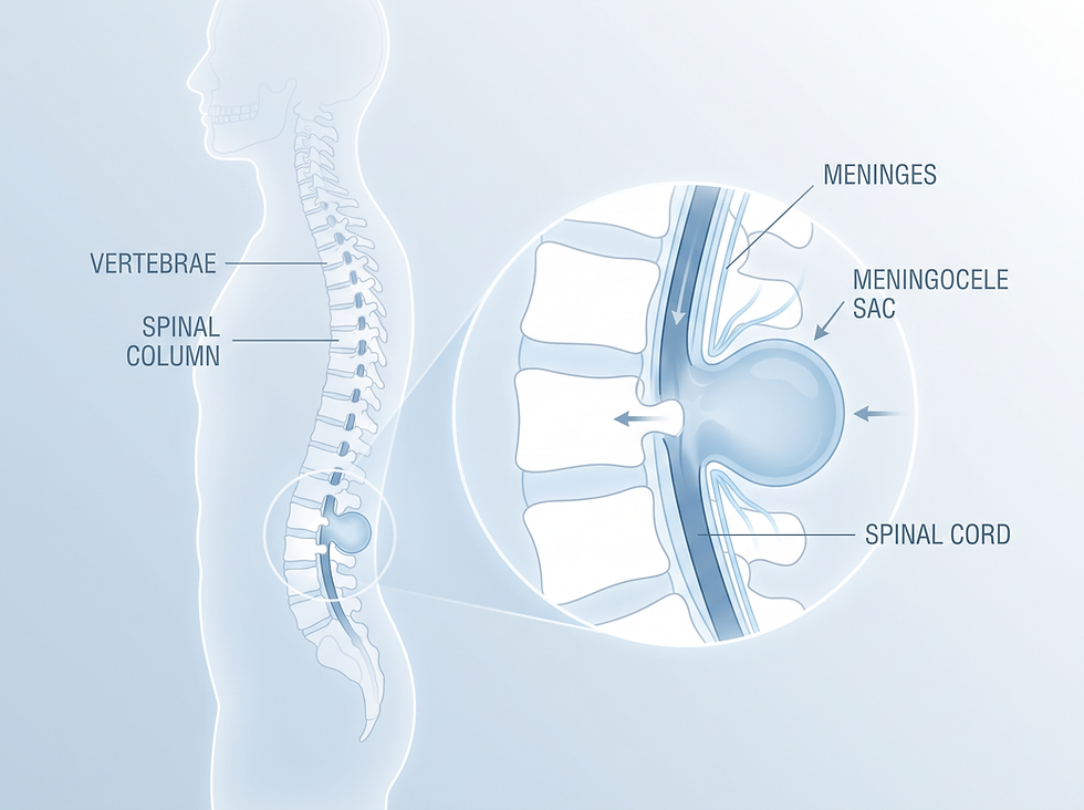
Healthcare providers categorize this condition into two primary types based on the presence of skin coverage over the protruding sac. This classification is critical for determining the urgency and specific approach of the surgical repair required after delivery.
Type of Meningocele | Description |
|---|---|
Open Meningocele | The protruding sac is not covered by skin, leaving the membranes exposed. |
Closed Meningocele | The protruding sac is completely covered by a layer of skin. |
Symptoms and Clinical Signs
The most prominent sign of a meningocele is the presence of a fluid-filled lump or sac on the infant's back. While many infants do not exhibit additional symptoms immediately, the condition can sometimes be associated with secondary clinical signs that require monitoring by a neonatal care team.
Common signs and potential symptoms include:
A visible, fluid-filled sac, typically located on the lower spine.
Potential muscle weakness in the lower extremities.
Changes in the sound or quality of the infant's cry.
Occasional back pain or discomfort.
Respiratory signs, such as shortness of breath or difficulty breathing.
Feeding difficulties, including trouble swallowing (dysphagia).
Physical signs like a fluttering heartbeat or persistent coughing.

Causes and Risk Factors
Meningocele is caused by a developmental interruption where the bones of the spine do not close completely around the spinal cord. While the exact cause can vary, research indicates that certain maternal health factors and medications may increase the likelihood of a neural tube defect occurring during pregnancy.
Risk Factor Category | Details and Associations |
|---|---|
Maternal Health | Chronic conditions such as diabetes in the pregnant person. |
Medications | The use of certain antiseizure or anticonvulsant medications during pregnancy. |
Developmental | Failure of the lower neural tube to close properly during early fetal growth. |
Diagnosis and Treatment Pathways
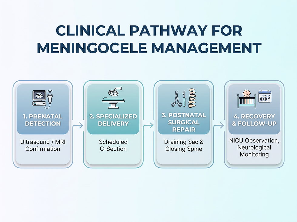
Diagnosis often begins during routine prenatal screenings through blood and urine tests. If a meningocele is suspected, advanced imaging such as ultrasound or MRI is used to confirm the diagnosis and assess the structure of the spine and spinal cord. In cases where the condition is detected before birth, a cesarean section (C-section) is often recommended to protect the sac from damage during delivery.
The primary treatment for meningocele is surgical repair, which typically occurs within the first few days of life. The goal of surgery is to drain the fluid-filled sac, reposition the meninges back into the spinal column, and close the opening in the spine to prevent infection and further complications.
Surgical Timing and Recovery
The timing of the surgical intervention depends on whether the meningocele is open or closed. Open meningoceles are typically repaired within 24 to 48 hours after birth to minimize the risk of infection. Closed meningoceles may be scheduled within 72 hours. Following the procedure, newborns usually remain in the hospital for up to two weeks to ensure proper healing and monitor for potential complications such as bleeding or increased intracranial pressure.
Outlook and Long-Term Care
The prognosis for children born with meningocele is generally excellent. Because the spinal cord is usually unaffected, long-term neurological issues are rare. However, regular follow-up appointments with pediatricians and specialists are essential to monitor growth, development, and the rare possibility of complications like a tethered spinal cord.
Conclusion
A meningocele diagnosis can be overwhelming for families, but modern medical interventions provide highly successful outcomes. With prompt surgical care and consistent follow-up, most children born with this condition go on to lead healthy, active lives without significant medical limitations.
Call to Action
If you are managing a prenatal diagnosis or caring for a newborn with a spinal sac, work closely with a multidisciplinary neonatal team. Early intervention and specialized neurosurgical care are the most effective ways to ensure a successful recovery and long-term wellness for your child.
Related reading: Fetal Surgery: Advanced Prenatal Interventions and Care
Frequently Asked Questions
1. What is the main difference between meningocele and myelomeningocele?
In a meningocele, only the protective membranes (meninges) protrude through the spine, while the spinal cord remains in place. In myelomeningocele, both the membranes and the spinal cord protrude, which typically causes more severe symptoms.
2. Is meningocele a common condition?
No, it is the least common type of spina bifida.
3. Can meningocele be detected before the baby is born?
Yes, it can often be identified during routine prenatal blood tests, urine tests, or ultrasound screenings.
4. Why is a C-section often recommended for babies with meningocele?
A C-section helps prevent physical trauma or rupture of the fluid-filled sac that could occur during a vaginal delivery.
5. How soon after birth is surgery performed?
Surgery is usually performed within 24 to 72 hours after birth, depending on whether the sac is open or covered by skin.
6. What is inside the meningocele sac?
The sac contains the meninges and cerebrospinal fluid (CSF), which is the fluid that normally cushions the brain and spinal cord.
7. Will my child have paralysis from a meningocele?
Paralysis is rare with this condition because the spinal cord itself is usually not damaged or displaced.
8. Are there long-term complications associated with meningocele?
While rare, complications can include increased pressure in the brain, infection, or a tethered spinal cord. Most children recover fully.
9. What are the risks of the surgical repair?
As with any surgery, risks include bleeding, infection, and potential reactions to general anesthesia.
10. How long is the hospital stay after surgery?
Most newborns stay in the hospital for approximately two weeks following the procedure to ensure proper healing.
11. Does maternal diabetes increase the risk of meningocele?
Yes, research suggests that chronic conditions like diabetes in the pregnant person can increase the risk of neural tube defects.
12. Can medications cause this condition?
Certain antiseizure medications taken during pregnancy have been linked to an increased risk of spina bifida variants like meningocele.
13. What is a tethered spinal cord?
This is a rare complication where the spinal cord becomes attached to the surrounding tissues, which can cause issues as the child grows.
14. Will my child need special therapy after surgery?
Most children do not require extensive long-term therapy, but regular check-ups are necessary to monitor their development.
15. Is meningocele considered a life-threatening condition?
With prompt surgical treatment, it is not typically life-threatening, and the outlook for a normal lifespan is excellent.
Medical Disclaimer: The information provided in this article is for educational purposes only and should not be used as a substitute for professional medical advice, diagnosis, or treatment. Always seek the advice of a qualified healthcare provider with any questions you may have regarding a medical condition.
Comments