Morning Glory Syndrome: Causes, Symptoms, and Management

Updated: 2 days ago
Medically reviewed by Dr. Baraa Alnahhal, MD · Last reviewed: September 2026
TL;DR: Morning Glory Syndrome (MGS) is a rare developmental condition where the back of the eye forms a funnel shape instead of a globe. It often leads to significant vision challenges and a high risk of retinal detachment. While the structural differences are permanent, symptoms and complications are manageable through specialized eye care.
Quick answer: Morning Glory Syndrome occurs when the optic disk and cup develop too wide and deep, creating a funnel-like appearance inside the eye. This rare condition typically affects one eye and is usually diagnosed before age two. While there is no direct cure for the structural differences, treatment focuses on managing complications like retinal detachment and nearsightedness to preserve as much vision as possible.
Overview of Morning Glory Syndrome
Morning Glory Syndrome is a rare congenital condition characterized by the incorrect development of the back of the eye. In a typical eye, the internal structure is globe-shaped; however, in those with this condition, the posterior segment takes on a funnel-like appearance. The name is derived from the visual resemblance of the optic disk to a morning glory flower when viewed during a medical examination.
This condition is almost always identified early in life, frequently before a child reaches two years of age. While it usually affects only one eye, it can occasionally present in both. When the structural differences occur without accompanying symptoms, medical experts refer to the condition as a morning glory disk anomaly. However, when these differences lead to symptoms or other health issues, it is classified as Morning Glory Syndrome.

Symptoms and Common Indicators
Morning Glory Syndrome is twice as likely to affect the left eye as the right eye. The symptoms can vary significantly between individuals, and not every person will experience all possible indicators. The most frequently reported symptoms involve changes in vision and the physical appearance of the eye:
Nearsightedness: Difficulty seeing objects at a distance (myopia).
Visual field gaps: Blind spots (scotomas) or an enlarged natural blind spot.
Crossed eyes: Misalignment of the eyes, known as strabismus.
Leukocoria: A pupil that appears white, silvery, yellowish, or gray.
In addition to primary eye symptoms, Morning Glory Syndrome can occur alongside other conditions. These may include neurological or developmental issues such as encephalocele, Moyamoya disease, and PHACE syndrome. Other associated conditions include neurofibromatosis type 2, Aicardi syndrome, and Chiari malformation type I.
Potential Causes and Risk Factors
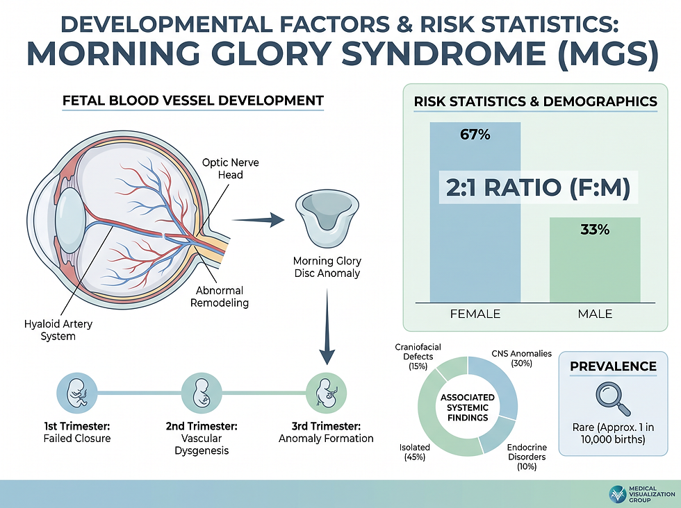
The exact mechanism behind the development of Morning Glory Syndrome is not fully understood, partly due to its rarity. However, it is known that the condition stems from disruptions in eye development that occur before birth. These disruptions appear to involve the underdevelopment of blood vessels supplying the eyes or issues with the development of the optic nerve itself.
One theory suggests that small blood clots (microemboli) in a developing fetus may play a role. These clots can potentially travel through openings in the heart and block blood flow to developing brain and eye tissues. This theory is supported by the fact that the left eye is more frequently affected, aligning with fetal blood flow patterns. While the condition can be associated with certain genetic syndromes, it is not typically considered a directly inheritable disorder.
Diagnosis and Medical Evaluation
Diagnosing Morning Glory Syndrome requires a comprehensive evaluation by an eye care specialist, particularly a retina specialist. The process involves a combination of physical examinations, vision testing, and advanced imaging of the internal structures of the eye.
Vision testing: Standard exams to check visual acuity and the extent of the visual field.
Slit lamp exam: A detailed inspection of the front and back of the eye using a specialized microscope.
Retinal imaging: Advanced scans like optical coherence tomography (OCT) and fluorescein angiography to capture detailed images of the retina.
Neurological imaging: Magnetic resonance imaging (MRI) may be used to check for associated brain or blood vessel conditions.
Management and Treatment Strategies
Because the structural differences in the eye are established before birth, there is no cure for Morning Glory Syndrome. Treatment is not directed at the condition itself but rather at managing the symptoms and preventing or treating complications. The primary goal of care is to preserve existing vision and improve the patient's quality of life.
Repair complications: Surgery to reattach a detached retina.
Manage symptoms: Corrective lenses for nearsightedness.
Prevent vision loss: Using an eye patch to treat or prevent lazy eye (amblyopia).
Ongoing monitoring: Regular check-ups to watch for new blood vessel growth or cataracts.

Outlook and Long-Term Prognosis
Morning Glory Syndrome is a lifelong condition that can significantly impact vision. While it is not life-threatening, the majority of individuals with the condition meet the criteria for being partially sighted or legally blind. Nearly 40% of people with the condition will experience a retinal detachment at some point in their lives, making regular monitoring essential.
For those with one-sided involvement, it is crucial to monitor the unaffected eye as well. The healthy eye may face an increased risk of cataracts or retinal detachment due to shared risk factors like nearsightedness. Early diagnosis and consistent follow-up care are the most effective ways to manage the challenges associated with this rare condition.
Take Action
If you or your child are experiencing vision changes, blind spots, or unusual pupil coloration, it is important to seek a comprehensive eye examination. Early intervention can help manage complications and provide the necessary support for living with vision impairment. Consult with an eye care specialist to develop a personalized management plan.
Frequently Asked Questions
Is Morning Glory Syndrome curable?
No, the structural differences in the eye are permanent and cannot be cured, but symptoms are manageable.
How did the condition get its name?
The optic disk resembles a morning glory flower when viewed during a medical exam.
Is it always diagnosed at birth?
Most cases are diagnosed in early childhood, typically before the age of two.
Can it affect both eyes?
It usually affects only one eye, but in rare cases, it can occur in both.
Is Morning Glory Syndrome life-threatening?
No, it is a vision-related condition and does not impact overall life expectancy.
What is the most common complication?
Retinal detachment is the most frequent complication, affecting about 40% of patients.
Does it run in families?
While there are rare records of multiple family members having it, it is not typically considered inheritable.
What is a morning glory disk anomaly?
This term is used when the structural differences are present but do not cause symptoms.
Can surgery fix the funnel shape?
No, surgery cannot change the fundamental shape of the eye's interior.
Who is most likely to be affected?
It is more common in females, with a 2:1 ratio compared to males.
Does it affect all ethnicities equally?
Research suggests it is less likely to affect Black individuals.
What vision level can I expect?
Most people meet the criteria for being partially sighted or legally blind, though some have near-normal vision.
Why is the left eye more commonly affected?
It is believed to be related to fetal blood flow patterns and the heart's development.
What associated conditions should I watch for?
Neurological issues like Moyamoya disease or PHACE syndrome can sometimes occur alongside MGS.
How often is monitoring needed?
Regular lifelong check-ups are necessary to watch for complications like retinal detachment or cataracts.
Medical disclaimer: The information provided in this article is for educational purposes only and is not intended as a substitute for professional medical advice, diagnosis, or treatment. Always seek the advice of your physician or other qualified health provider with any questions you may have regarding a medical condition.
Source date: December 8, 2023. This article is current based on the most recent available data from the primary source.
Comments