Cavernous Malformations: The "Mulberry" Clusters of Blood Vessels in the Brain, Explained

Updated: 2 days ago
Medically reviewed by Dr. Baraa Alnahhal, MD · Last reviewed: September 2026
Editorial note: This article is for educational purposes only and is not medical advice. It is based on current clinical guidance (updated July 1, 2026) and should not replace a consultation with a qualified healthcare professional.
Quick Answer
A cavernous malformation is a small cluster of abnormal, slow-flowing blood vessels in the brain or spinal cord, often described as looking like a mulberry. Most cause no symptoms, but they can leak blood and trigger seizures, headaches, or stroke-like problems. About 20% are inherited through genes known as CCM1, CCM2, and CCM3. Treatment ranges from careful monitoring to medication to surgical removal.
What is a cavernous malformation?
A cerebral cavernous malformation (CCM) is a small cluster of abnormal blood vessels in the brain or spinal cord. The vessels contain slow-moving blood that is usually clotted. They look like small mulberries and have thin walls that may leak blood.
CCMs vary in size. Often they are less than 1/2 inch (1 centimeter). They are also called cavernomas or cavernous angiomas.

By the numbers, CCMs are more common than many people assume. About 1 in 100 to 200 people have at least one cavernous malformation, and they probably form before or shortly after birth. Population estimates put overall prevalence between 1 in 200 and 1 in 1,000 people, with the familial (inherited) form accounting for about 20% of cases.
Key facts at a glance
How common CCMs are: about 1 in 100 to 200 people have one (Johns Hopkins Medicine)
Prevalence range: 1 in 200 to 1 in 1,000 people overall (Orphanet)
Share that are inherited (familial): about 20% (Mayo Clinic)
Typical size: usually less than 1/2 inch (1 cm) (Mayo Clinic)
People with brain CCMs who never have symptoms: about 25% (Johns Hopkins Medicine)
Most common first symptom: seizure, in about 50% of people diagnosed (Alliance to Cure Cavernous Malformation)
What are the symptoms of a cavernous malformation?
Most cerebral cavernous malformations do not cause symptoms at all. In fact, about a quarter of people with cavernous malformations in the brain never develop symptoms. When symptoms do occur, they depend heavily on where the malformation sits.
If a CCM is in the brain, it can sometimes lead to seizures or other bleeding-related symptoms such as weakness, trouble speaking, or changes in vision. If a CCM is in the spinal cord, symptoms can include trouble with the bowel and bladder or difficulty moving or feeling the legs and sometimes the arms.
Seizures — the most common first symptom; about half of diagnosed patients present this way
Hemorrhagic stroke — a stroke caused by bleeding in the brain
Bad headaches — described as unusually severe
Weakness — in the arms or legs
Numbness — loss of feeling, often on one side
Trouble speaking — difficulty forming or understanding speech
Poor memory and attention — cognitive changes that develop gradually
Balance and walking trouble — unsteadiness, difficulty walking
Vision changes — such as double vision
Bowel and bladder symptoms — with spinal cord malformations
One point bears emphasis: symptoms can get worse over time with repeated bleeding. Bleeding can happen again soon after the first bleed or much later. In some people, a repeat bleed may never occur.
What is a cavernous malformation hemorrhage, and how risky is it?
The most serious complication of a CCM is bleeding into the brain or spinal cord, called a hemorrhage. Brain hemorrhages can cause many symptoms, such as seizures.
The risk numbers vary enormously depending on one key fact: whether the malformation has bled before and where it sits.
Incidental CCM (found on imaging, never caused symptoms): about 0.08% per patient-year — very, very low
First symptomatic hemorrhage (any patient): about 0.4% to 2.4% per year
After a first symptomatic hemorrhage (rebleed): 3.8% to 29.5% per year
Within 5 years of a first bleed: about 42% re-hemorrhage risk
Brainstem location: higher risk than other locations
The incidental-lesion numbers come from a study that tracked 107 patients with incidental lesions for 1,311 patient-years over more than 10 calendar years — only two hemorrhages were recorded. That is why doctors frequently recommend watchful waiting for silent malformations.
What causes cavernous malformations?
Most cerebral cavernous malformations happen by chance. This is called the sporadic form — there is only one malformation without any family history. The sporadic form often happens alongside another irregular vein that looks like a witch's broom, called a developmental venous anomaly (DVA).
About 20% of people with a CCM have a genetic form passed down in families, known as familial cavernous malformation syndrome. Familial CCM is inherited in an autosomal dominant pattern, meaning a person needs only one copy of the affected gene from either parent to have the condition.
Radiation to the brain or spinal cord also may result in CCMs within 2 to 20 years afterward. Other rare syndromes may be associated with CCM.
CCMs compared with other brain vascular malformations
Cavernous malformation (CCM) — slow-flowing, clotted-blood mulberry cluster with thin walls; usually under 1 cm
Arteriovenous malformation (AVM) — tangled connection of arteries and veins
Dural arteriovenous fistula — abnormal connection in the dura (the tissue covering the brain)
Developmental venous anomaly (DVA) — irregular vein with a witch's broom appearance; commonly co-occurs with sporadic CCMs
Capillary telangiectasia — dilated capillaries in the brain tissue
What genes are linked to familial cavernous malformations?
To date, research has identified three genetic changes responsible for cavernous malformations passed down through families. Almost all familial cases have been traced through those genetic changes:
KRIT1 (also known as CCM1)
MGC4607 (also known as CCM2)
PDCD10 (also known as CCM3)
These genes affect the leakiness of blood vessels and the proteins that keep the blood vessel cells together. When genetic variations affect protein production, cell junctions weaken and CCMs can form.
Familial CCMs change over time. They may increase in number or increase or decrease in size. The most serious form of the disease is usually seen in people who have changes in the CCM3 gene. People with CCM3 changes may have symptoms at a younger age, more lesions, and a greater risk of hemorrhage.
Who is at risk for cavernous malformations?
Most CCMs have no clear cause, and the exact cause is unknown. That is also why CCMs cannot be prevented — neither the sporadic form, which happens by chance, nor the genes that cause familial CCMs can be modified.
Family history of CCM — inherited form; person may have multiple malformations
A mutation in CCM1, CCM2, or CCM3 — causes familial cavernous malformation syndrome
Prior radiation to the brain or spinal cord — may result in CCMs 2 to 20 years afterward
Certain rare syndromes — associated with CCM in some patients
Most people who develop symptoms are between 20 and 50 years old. Seizures tend to get worse and happen more often as people with CCMs get older.
How is a cavernous malformation diagnosed?
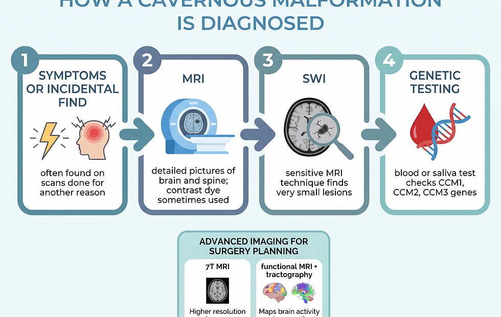
Often people with CCMs have no symptoms at all, and the malformation is found during brain imaging done for another condition. Sometimes specific symptoms — a seizure, an unusual headache — prompt the search.
MRI — a detailed picture of the brain or spine; a contrast dye is sometimes injected into a vein
SWI (susceptibility-weighted imaging) — a more sensitive MRI technique used to find very small lesions, common in familial CCMs
Genetic testing — blood or saliva test that finds the CCM1, CCM2, or CCM3 gene changes; paired with genetic counseling for people with a family history
7-tesla MRI (7T) — advanced imaging that distinguishes CCMs from other types of brain disease and aids treatment planning
Functional MRI — measures blood flow in the active parts of the brain, useful when surgery is being planned
Tractography — creates a map of the brain's wiring to make surgery as safe as possible
Genetic testing is often recommended for people who have MRI evidence of multiple CCMs without a DVA, or a family history of CCMs. Because CCMs can change in size and number over time, people usually need lifelong monitoring with regular MRIs.
What treatments work for cavernous malformations?
CCMs are treated by specialists, including neurologists, cerebrovascular neurologists, neurosurgeons, and neuroradiologists. There is no standard treatment for all CCMs; the plan depends on the malformation’s size, location, symptoms, and bleeding history.
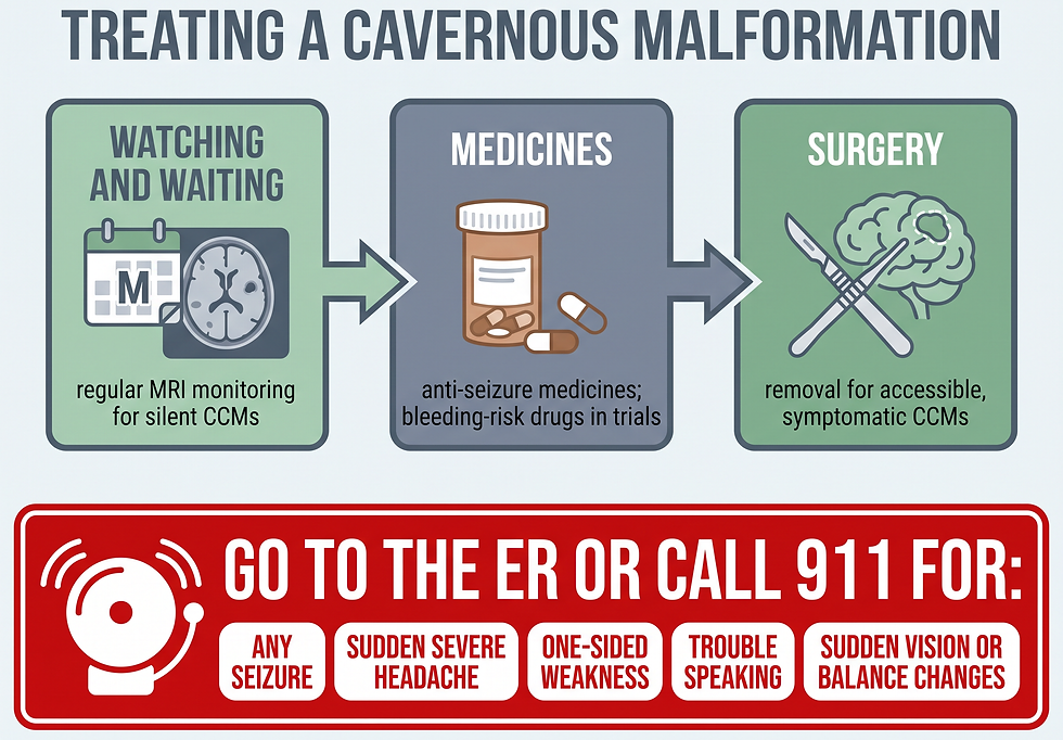
Watching and waiting — for silent, incidental CCMs with very low bleed risk; close monitoring with regular MRI imaging
Medicines — for seizures related to a CCM; anti-seizure medicines, plus research exploring medicines that could reduce bleeding risk
Surgery — for accessible CCMs causing seizures, repeated bleeds, or progressive symptoms; removing or treating the malformation surgically
If the treatment plan includes surgery, advanced imaging technologies may be used — functional MRI to measure blood flow in active brain areas, and tractography to map the brain and make surgery as safe as possible.
Surgery is generally not recommended if you are asymptomatic, especially if the CCM is in a deep or hard-to-reach location such as the brainstem. If you have had a symptomatic hemorrhage in a location that is easily accessible, surgery may be recommended.
Medicines that reduce the chance of further bleeding — rather than surgery — are being tested in clinical trials. Talk to a healthcare professional about which clinical trials may be available. Researchers are also studying advanced imaging (quantitative susceptibility mapping and dynamic contrast-enhanced permeability imaging) to better predict how the disease will progress.
What should you do in an emergency?
Seek medical help right away if you have any symptoms of a seizure. Also get medical help right away for symptoms suggesting a CCM or brain bleeding — seizure, severe headache, numbness, vomiting with headache, or physical weakness.
Go immediately to the emergency room or call 911 for these warning signs:
Seizure — any seizure episode, including a first-ever seizure
Sudden severe headache — often described as "the worst headache of my life," sometimes with vomiting
Sudden weakness — in the face, arm, or leg, often on one side
Sudden numbness — loss of feeling, especially on one side
Trouble speaking — difficulty speaking or understanding speech
Vision changes — sudden double vision or loss of vision
Balance problems — sudden difficulty walking or staying steady
What should you expect if you see a doctor?
If a CCM is suspected, a referral to an experienced specialist — a neurologist or neurosurgeon with cerebrovascular expertise — provides the best outcome.
Before the appointment, keep a detailed symptoms calendar noting the time, what happened, and how long it lasted. Bring a list of all medicines, vitamins, and supplements, along with any recent brain scans. It is common not to remember everything during a seizure, so a witness’s account is valuable.
In the meantime, certain conditions and activities can trigger seizures. It may help to avoid alcohol, avoid nicotine, get enough sleep, and reduce stress.
Useful questions to ask include: Do I have one or more cavernous malformations? Where are they located? What do those areas of my brain do? Should I have genetic tests, and should my children or other family members be tested? How often do I need follow-up imaging? And for surgery: how long does it take, how long is recovery, and how many CCMs has this surgeon treated?
What is the bottom line?
Cavernous malformations are mulberry-shaped clusters of thin-walled blood vessels in the brain or spinal cord. They are surprisingly common — roughly 1 in 100 to 200 people have one — yet most never cause a symptom.
The practical plan is simple: know the warning signs (seizure, severe headache, one-sided weakness), act immediately if they appear, and see a specialist early if you have a family history, since about 20% of cases are inherited and run in families. For silent malformations, careful monitoring with MRI is often the best course. For malformations that bleed or cause seizures, medication and, in accessible locations, surgery can change the trajectory.
Know the warning signs. Share this guide with family members — especially if CCMs run in your family — and bring it to your next specialist appointment.
Frequently asked questions
What is a cavernous malformation?
A cavernous malformation is a small cluster of abnormal, slow-flowing blood vessels in the brain or spinal cord, shaped like a mulberry and usually under 1 centimeter. Its thin walls may leak blood. Most cause no symptoms.
Are cavernous malformations dangerous?
Usually not. About a quarter of people with brain CCMs never develop symptoms, and incidental (never-bleeding) lesions have a very low hemorrhage rate of about 0.08% per patient-year. Risk rises after a first bleed and with brainstem location.
Can a cavernous malformation cause seizures?
Yes — seizure is the most common first symptom. Roughly half of people diagnosed with a CCM experience a seizure as their first sign, and seizures tend to become more frequent with age.
Are cavernous malformations genetic?
About 20% are. The familial form is inherited in an autosomal dominant pattern through one of three genes: KRIT1 (CCM1), MGC4607 (CCM2), or PDCD10 (CCM3). People with familial CCM usually have more than one malformation.
Can you prevent cavernous malformations?
No. CCMs cannot be prevented because the exact cause is unknown and the genes behind the familial form are passed down through families.
How are cavernous malformations diagnosed?
Usually with MRI, which can show the malformation and its location. A more sensitive technique called susceptibility-weighted imaging (SWI) finds very small lesions. Genetic testing (blood or saliva) confirms the familial form.
Do cavernous malformations always need surgery?
No. Many are managed with careful monitoring and regular MRI. Surgery is considered for malformations that cause seizures, repeated bleeds, or progressive symptoms — especially in locations where removal is safe.
When should I go to the emergency room?
Go immediately or call 911 for any seizure, a sudden severe headache (especially with vomiting), sudden one-sided weakness or numbness, trouble speaking, or sudden vision or balance problems.
References
Mayo Clinic — Cavernous malformations: Symptoms & causes (updated July 1, 2026). mayoclinic.org/diseases-conditions/cavernous-malformations/symptoms-causes/syc-20360941
Mayo Clinic — Cavernous malformations: Diagnosis & treatment (updated July 1, 2026). mayoclinic.org/diseases-conditions/cavernous-malformations/diagnosis-treatment/drc-20360942
National Institute of Neurological Disorders and Stroke — Cerebral Cavernous Malformations Information Page. ninds.nih.gov/health-information/disorders/cerebral-cavernous-malformations
Johns Hopkins Medicine — Cavernous Malformations. hopkinsmedicine.org/health/conditions-and-diseases/cavernous-malformations
Orphanet — Familial cerebral cavernous malformation. orpha.net/en/disease/detail/221061
Alliance to Cure Cavernous Malformation — Hemorrhage (and Seizure). alliancetocure.org/home/cavernous-angioma-in-depth/symptoms-and-hemorrhage/hemorrhage/
Sandmann ACA, et al. Incidence of cerebral cavernous malformations among people in Germany, Sweden and the UK: a population-based study. Journal of Neurology, Neurosurgery & Psychiatry. 2026. jnnp.bmj.com/content/97/9/790
Mayo Clinic — Genetic testing. mayoclinic.org/tests-procedures/genetic-testing/about/pac-20384827
Mayo Clinic — MRI (magnetic resonance imaging). mayoclinic.org/tests-procedures/mri/about/pac-20384768
Goldstein HE, et al. Epidemiology of cavernous malformations. Current Treatment Options in Cardiovascular Medicine. 2017. pubmed.ncbi.nlm.nih.gov/28552146/
Comments