Angiokeratoma Explained: A Benign Skin Bump That Is Often Mistaken for Something Worse

Updated: 2 days ago
Medically reviewed by Dr. Baraa Alnahhal, MD · Last reviewed: September 2026
Editorial note: This article is for general education only. It is not medical advice, a diagnosis, or a treatment plan. Angiokeratomas are benign (noncancerous) skin lesions — but because some can look like other conditions, including melanoma, any new, changing, bleeding, or unusual skin lesion deserves evaluation by a dermatologist or other healthcare provider. If you have many angiokeratomas appearing in the "swimsuit" area, a medical evaluation is important to rule out an underlying metabolic condition.
TL;DR
Angiokeratoma is a benign (noncancerous) skin lesion caused by small blood vessels (capillaries) that widen or break open near the skin's surface while the overlying skin thickens, forming firm red, purple, or black bumps [1]. It is not cancer and not contagious [1]. There are four main types — the most common affects the scrotum or vulva after age 50, and one type signals an inherited metabolic disorder called Fabry disease [1] [7]. Most angiokeratomas never need treatment, but bleeding, irritated, or bothersome lesions can be removed with cryotherapy, laser, or minor surgery [1].
Quick Answer: What Is Angiokeratoma?
Angiokeratoma is a harmless skin bump that forms when a tiny blood vessel near the skin's surface enlarges and the skin above it thickens, creating a firm, rough, dome-shaped bump that is red, blue, purple, or black [1]. It is a benign vascular lesion — not cancer, not an infection, and not a sexually transmitted disease [1]. The four main types are Fordyce (genitals), Mibelli (hands and toes), circumscriptum (present at birth), and corporis diffusum (linked to Fabry disease) [1] [4]. Treatment is optional and usually reserved for lesions that bleed, itch, or bother you cosmetically.
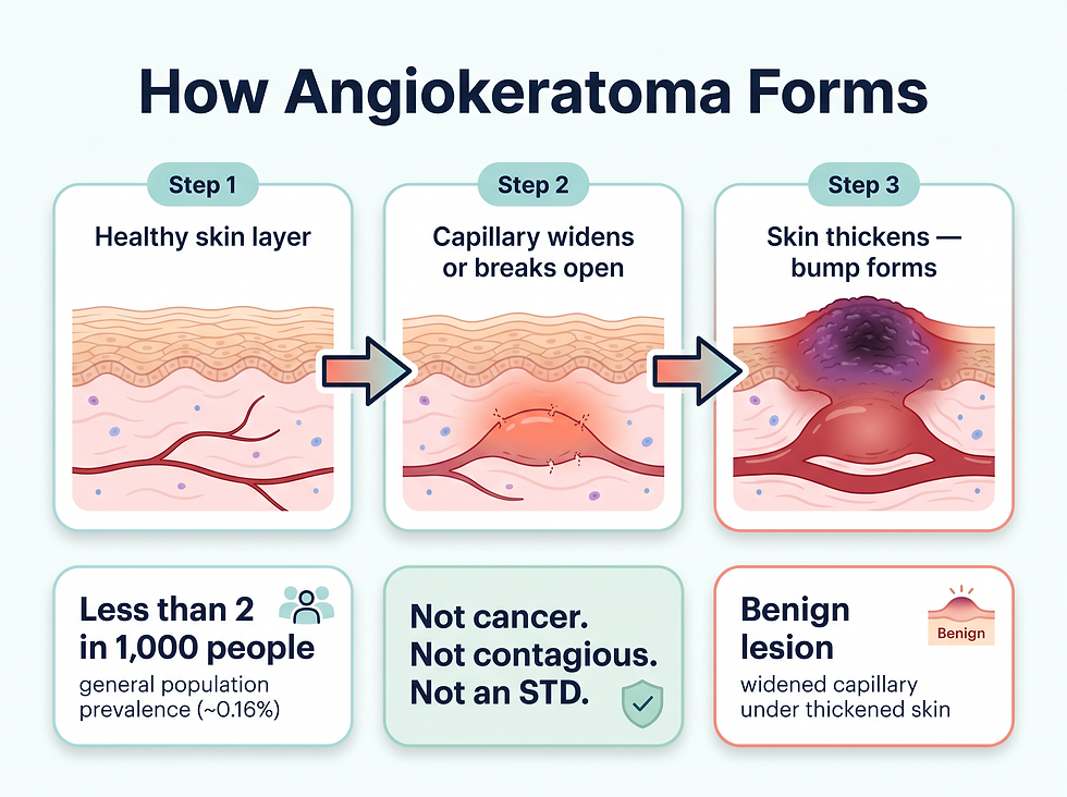
A dilated capillary (tiny blood vessel) swells and pushes upward into thickened skin, forming a firm, dome-shaped red-to-black bump. About 2 in 1,000 people are affected, the solitary type accounts for 80% of angiokeratomas, and the condition is not cancer and not contagious.
What Exactly Is Angiokeratoma?
Angiokeratomas are hard bumps on your skin. The word itself tells you what the lesion is made of — in Greek, "angio" means vessels, "kera" means horn, and "toma" means tumor [1]. Put simply, an angiokeratoma is a tiny tumor of thickened skin sitting on top of a widened blood vessel.
The condition develops when the tissues that support the walls of your capillaries — the smallest blood vessels in your body, so tiny that you need a microscope to see them — lose their elasticity [1]. The body has roughly 10 billion capillaries, and they connect arteries to veins so oxygen and nutrients can reach every cell [1]. When a capillary's supporting tissue weakens, the vessel swells and can push into the skin above it, forming a raised, discolored bump called a papule [1]. Meanwhile, the overlying skin thickens and takes on a rough, pebble-like or scaly texture [1].
Importantly, angiokeratomas are a type of benign (noncancerous) tumor [1]. They are not skin cancer, they are not contagious, and having them does not raise your cancer risk [1].
Term you might hear | What it means |
Angiokeratoma | A hard, discolored skin bump formed by a widened capillary under thickened skin [1] |
Capillary | The smallest blood vessel in the body, connecting arteries and veins [1] |
Benign tumor | A noncancerous growth that does not spread or invade other tissues [1] |
Papule | A small, raised bump on the skin [1] |
Dermis | The middle layer of skin, where the widened vessels sit [1] |
Epidermis | The top layer of skin, which thickens over the lesion [1] |
How Common Are Angiokeratomas?
Angiokeratomas are rare in the general population. Overall prevalence is estimated at about 0.16% of Americans — fewer than 2 in 1,000 people [1]. Independent dermatology references place the figure at roughly 0.1% to 0.2% of the general population, consistent with the same classic study [5].
That overall number, however, understates how common certain types are in specific groups. In a 2019 prevalence study of 213 white/European American adults who underwent genital examination during skin-cancer screening, genital angiokeratomas were found in 30% of adults — and they were strongly associated with being male (about 2.4 times more likely) and being older than 50 (about 3.4 times more likely) [3].
Subtype | Share of all angiokeratomas | Who it typically affects |
Solitary angiokeratoma | ~80% | Adults, usually on the lower limbs; can occur anywhere [5] |
Fordyce (genital) | ~15% | Adults; most common over 50; more frequent in white men [1] [5] |
Mibelli | ~3% | Adolescent girls and young women, on hands and toes [4] [5] |
Circumscriptum | Rare | Present at birth; the only type more common in females [1] [5] |
Corporis diffusum | <1% | People with lysosomal storage diseases such as Fabry disease [5] [7] |
The solitary form is by far the most common presentation — about 83% of studied patients had a single lesion [8] — while a minority of people develop many bumps, sometimes even hundreds [1].
What Are the Four Types of Angiokeratoma?
There are four recognized types, each with its own typical location, age of onset, and underlying cause [1] [4] [5].
Angiokeratoma of Fordyce is the best-known type. It appears on the scrotum or penis and, less often, on the vulva — which is why it is also called angiokeratoma of the scrotum or of the vulva [1]. It most commonly affects people aged 50 and over, and in the United States it occurs mostly in white men [1] [3]. Some people develop many lesions, even hundreds [1]. Pressure from conditions such as varicoceles (swollen scrotal veins), inguinal hernias, or pregnancy can contribute [1].
Angiokeratoma of Mibelli most commonly affects adolescent girls and young women [4]. Lesions typically appear on the webbing between the fingers and toes, and may also form on the knees, elbows, and breasts [1]. These lesions tend to bleed easily, and are sometimes preceded by a history of chilblains (cold-related skin inflammation) [4].
Angiokeratoma circumscriptum is present when a baby is born as flat, discolored lesions that gradually turn into raised, red, scaly patches that may bleed [1]. The plaques are usually smaller than half an inch and most commonly appear on one side of the buttock or thigh; in rare cases they may cover a quarter of the body [1]. This is the rarest type and the only one more common in females [1].
Angiokeratoma corporis diffusum is the most medically important type because it occurs in people with lysosomal storage diseases, most notably Fabry disease — an inherited metabolic disorder in which the body cannot properly break down certain waste products [1] [7]. Lesions show up on the genitals, abdomen, and the "swimming trunk" area between the hips and knees, and may appear during childhood, adolescence, or adulthood [1]. In people with Fabry disease, angiokeratomas affect roughly 83% of males and 80% of females and can be the earliest visible sign of the condition [7].
Type | Typical location | Typical age | Key features |
Fordyce | Scrotum, penis, vulva | 50+, sometimes younger | Most common type; can number in the hundreds; linked to varicocele, hernia, pregnancy pressure [1] |
Mibelli | Fingers, toes, knees, elbows, breasts | Adolescents and young women | Bleeds easily; often preceded by chilblains [1] [4] |
Circumscriptum | Buttock, thigh (one side) | Present at birth | Flat patches become raised scaly plaques; can spread; rare [1] |
Corporis diffusum | Genitals, abdomen, "swimming trunk" area | Childhood to adulthood | Signals an underlying metabolic disease (Fabry); ~83% of males and ~80% of females with Fabry have lesions [1] [7] |
What Causes Angiokeratoma?
Angiokeratomas occur when the tissues that support capillary walls lose their elasticity [1]. The weakened, stiffened capillaries swell and may break open, pushing into the skin to form the raised bump [1]. Similar widening of surface veins is what happens with spider veins [1].
In many cases the cause is unknown or built in — some forms can be genetic [1]. Other recognized contributors include:
Proposed factor | How it contributes |
Genetics | Some forms run in families; corporis diffusum is tied to inherited metabolic disorders [1] [7] |
Vascular malformations | Abnormal vessel structure predisposes the capillary to widening [1] |
Pressure on blood vessels | Varicoceles and inguinal hernias press on scrotal capillaries; pregnancy pressure can cause vulvar lesions [1] |
Chronic irritation / trauma | A solitary angiokeratoma can develop at an injury site years after the trauma [1] |
Lysosomal storage disease | In Fabry disease, waste buildup weakens vessel walls; lesions appear in ~83% of affected males [7] |
It is worth emphasizing what angiokeratoma is not caused by. It is not caused by infection, poor hygiene, or sexual contact. It is not a sexually transmitted disease — you cannot catch it from anyone, and you cannot give it to anyone [1].
What Are the Symptoms of Angiokeratoma?
Angiokeratomas often start out resembling warts [1]. They can appear anywhere on the body, and the bumps typically begin red and then darken to purple or black as the overlying skin thickens [1]. They are less noticeable on darker skin [1].
One useful distinguishing feature: when you press on an angiokeratoma, it does not lighten (blanch) the way some other red skin spots do [1].
Typical angiokeratomas are:
Dome-shaped with a rounded top [1]
Firm or hard to the touch [1]
Red, blue, purple, or black in color [1]
Smaller than a quarter of an inch (5 millimeters) — usually 2 to 5 mm [1] [4]
Rough on the surface, with a pebble-like or scaly texture [1]
You may have one lesion (solitary angiokeratoma) or as many as 100 [1]. Most angiokeratomas cause no pain or symptoms at all — which is a large part of why they go unnoticed or get mistaken for something else [4].
Symptom | How it shows up |
Skin bumps | Firm, dome-shaped, rough, red-to-black papules under a quarter-inch wide [1] |
No blanching | The bump does not lighten when pressed [1] |
Bleeding | Some lesions bleed easily, even when slightly bumped [1] [5] |
Itching or burning | Irritation around the lesion; reported in a minority of patients [1] [5] |
Pain | Occasional mild pain; blood clots inside a lesion can be painful [1] [5] |
Appearance concern | Many people simply dislike how the bumps look [1] |
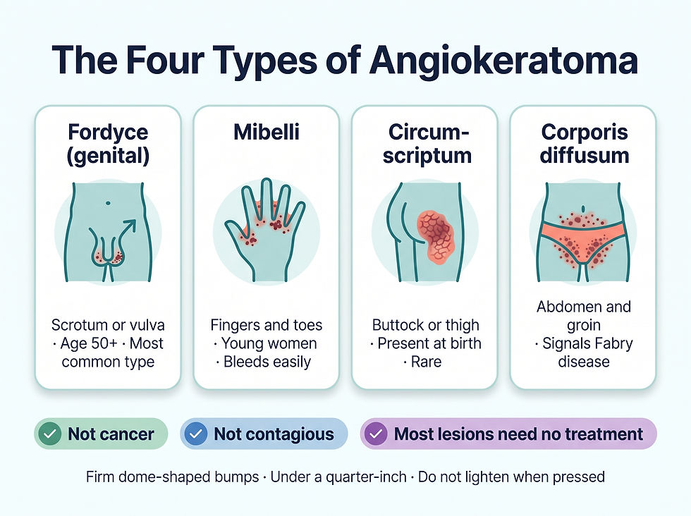
The four angiokeratoma types differ by location and appearance — genital, finger/toe, birthmark-like plaque, and widespread swimsuit-area lesions — but share reassuring features: not cancer, not contagious, not an STD, and most lesions need no treatment.
Is Angiokeratoma Dangerous?
For the vast majority of people, angiokeratoma is not dangerous. The lesions are benign, they do not turn into cancer, and having them does not increase your cancer risk [1]. The blood clots that can form inside a lesion are small, localized, and painful — but they are not the dangerous kind: they do not threaten your blood supply or raise the risk of stroke or heart attack [1].
The two realistic downsides are physical and emotional. Physically, some lesions bleed when bumped, and some itch or burn [1] [5]. Emotionally, the bumps — especially on the genitals — can make people feel self-conscious, and those feelings can contribute to depression or anxiety [1].
There is one important exception that warrants attention: widespread angiokeratomas (corporis diffusum) can be a visible sign of Fabry disease, a serious inherited metabolic disorder that, if untreated, can damage the kidneys, heart, and nervous system over time [1] [7]. If you have many small dark-red-to-black bumps across the lower abdomen, groin, and "swimsuit" area — particularly if they appeared in childhood or adolescence — tell your healthcare provider, as simple enzyme or genetic testing can rule this in or out [7].
How Is Angiokeratoma Diagnosed?
Diagnosis is usually straightforward and starts with a visual exam — often by a dermatologist, a doctor who specializes in skin conditions [1]. The challenge is that angiokeratomas can mimic other conditions. They can resemble warts or genital warts, and some lesions can mimic melanoma, the most dangerous form of skin cancer [1] [4].
To see the lesion clearly, your provider may use dermoscopy — a magnifying microscope device that lets them examine the bump's structure without touching it [1] [4]. If the diagnosis is in doubt, or if cancer needs to be ruled out, a skin biopsy removes a small sample for laboratory examination [1] [4].
Step | What happens | Why it matters |
Visual examination | Provider inspects the bump's color, texture, and location [1] | Most angiokeratomas are recognizable on sight [4] |
Dermoscopy | Magnified view of the lesion's structure [1] [4] | Helps distinguish it from melanoma and other lesions [4] |
Skin biopsy (if needed) | Small tissue sample sent to a lab [1] [4] | Confirms the diagnosis and rules out cancer [1] |
Further testing (rare) | Enzyme or genetic testing | Done when widespread lesions suggest Fabry disease [7] |
Because the genital form can look like genital warts, the diagnosis matters for peace of mind: it confirms you are dealing with something benign and not contagious — no treatment of a partner is ever needed [1].
Is There Treatment for Angiokeratoma?
Angiokeratomas do not go away on their own, and they can get bigger or multiply over time [1]. That said, most angiokeratomas never need treatment [1]. The decision to treat is usually about comfort and confidence rather than health: treatment makes sense if the lesions bleed, itch, cause pain, or bother you cosmetically [1].
All treatments are office procedures. Your provider may apply a local anesthetic to numb the skin, and you should know that some treatments can leave small scars [1].
Treatment | How it works | Best for |
Cryotherapy | Freezing temperatures destroy the lesion [1] | Small, accessible bumps |
Electrosurgery / fulguration | Heat destroys the lesion [1] | Bleeding-prone lesions |
Laser surgery (ablation) | High-intensity light removes the lesion [1] | Cosmetic results; genital lesions |
Skin excision | Surgical removal of the lesion and damaged capillary [1] | Solitary or stubborn lesions |
Newer studies continue to evaluate laser options, including long-pulsed alexandrite laser for scrotal lesions, which has shown good efficacy and safety in clinical study [11].
One honest caveat: treated lesions are unlikely to come back at the same spot, but new angiokeratomas can develop in other places on your body [1]. People with many lesions are more likely to have recurrences after treatment than people with one or a few [4].
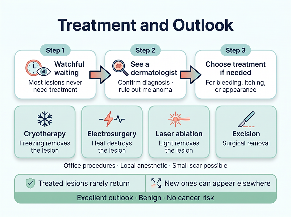
Care for angiokeratoma starts with watchful waiting for most lesions, followed by four removal options when needed — cryotherapy, electrosurgery, laser, or excision. Treated spots rarely return, though new ones can appear elsewhere, and the overall outlook is excellent since the condition is benign.
What Is the Outlook for Angiokeratoma?
The prognosis for angiokeratoma is excellent. These lesions are benign, they do not become cancerous, and no deaths or serious harm from the condition itself have been reported [1] [4].
The main issues are quality of life rather than health: self-consciousness about appearance (which can feed depression or anxiety in some people), occasional bleeding, and the small chance that a lesion mimics something more serious — which is exactly why a one-time diagnosis by a healthcare provider is worthwhile even when no treatment is planned [1].
If you live with genital angiokeratomas, a few practical points help. Avoid rough friction or scratching of the area, as some lesions bleed easily [1] [4]. And remember the reassurance that clinicians stress: the condition is common with age, it is benign, and it is not anything you caught [4].
Can Angiokeratoma Be Prevented?
Because angiokeratomas arise from natural changes in capillary support tissues, from genetics, or from pressure on blood vessels, there is no proven way to prevent them [1]. Maintaining general skin health — protecting skin from chronic irritation and injury, and promptly evaluating any new or changing skin lesion — is the best practical approach.
If you are pregnant or have a varicocele, know that these are recognized contributing factors for genital angiokeratomas [1], but they do not make them inevitable, and the lesions remain benign regardless.
Key Takeaways
Angiokeratoma is a benign, noncancerous, noncontagious skin bump formed when a tiny capillary widens or breaks open under thickened skin, producing a firm, rough, red-to-black bump usually smaller than a quarter inch [1]. Four types exist — Fordyce (genitals, most common after age 50), Mibelli (hands and toes), circumscriptum (present at birth), and corporis diffusum (linked to Fabry disease) [1] [4]. It affects roughly 0.16% of Americans, though genital forms are found in about 30% of some adult screening populations [1] [3]. It is not cancer, not an STD, and not dangerous [1]. Most lesions never need treatment; removal options include freezing, heat, laser, or excision for lesions that bleed or bother you [1].
If you have new, changing, bleeding, or dark skin bumps — especially on the genitals — see a dermatologist for confirmation. If you have many widespread bumps in the "swimsuit" area, ask about evaluation for Fabry disease.
Frequently Asked Questions
Is angiokeratoma cancer?
No. Angiokeratoma is a benign (noncancerous) lesion — a widened capillary under thickened skin. Having angiokeratomas does not increase your risk of skin cancer [1].
Is angiokeratoma an STD? Can I pass it to my partner?
No. Angiokeratomas are not a sexually transmitted disease or infection. You cannot catch them from anyone, and you cannot give them to anyone [1]. Genital forms can look like genital warts, which is why a provider may confirm the diagnosis — but they are entirely unrelated to sexual activity [1].
What causes angiokeratoma of Fordyce (scrotum)?
It develops when capillary support tissues lose elasticity and the vessels swell into the skin [1]. Contributing factors include varicoceles (swollen scrotal veins), inguinal hernias, and general pressure on the blood vessels [1]. It is most common in men over 50 [1] [3].
How common are angiokeratomas?
Overall prevalence is about 0.16% of Americans — fewer than 2 in 1,000 [1]. Genital forms are far more common in specific groups: one study found them in 30% of examined white/European American adults, with rates roughly 2.4 times higher in men and 3.4 times higher in adults over 50 [3].
Do angiokeratomas go away on their own?
No. They persist unless treated and can grow or multiply over time [1]. However, most never require treatment, and treated lesions are unlikely to return at the same spot (though new ones can appear elsewhere) [1].
When should I worry about angiokeratoma?
Angiokeratomas themselves are benign. Reasons to seek care include a lesion that changes, grows quickly, or bleeds repeatedly (a provider should confirm it is not melanoma), and — separately — the appearance of many widespread dark bumps in the "swimsuit" area, which can signal the inherited disorder Fabry disease [1] [7].
What are the treatment options?
Office procedures include cryotherapy (freezing), electrosurgery or fulguration (heat), laser ablation, and surgical excision, usually with local anesthetic [1]. Scarring is possible with some methods [1].
Can angiokeratoma mean I have Fabry disease?
Not usually — most angiokeratomas are isolated and benign. But the widespread form (angiokeratoma corporis diffusum) occurs in about 83% of males and 80% of females with Fabry disease and can be its earliest visible sign [7]. If you have many lesions in the groin and lower abdomen area, ask your provider about evaluation.
References
Cleveland Clinic — Angiokeratoma (medically reviewed, last updated 06/08/2022)
StatPearls — Angiokeratoma Circumscriptum (updated 07/20/2021)
Medscape — Angiokeratoma of the Scrotum (updated 06/07/2022)
Pathology Outlines — Angiokeratoma (last updated 01/15/2026)
Leung A.K.C., Barankin B. (2015) — Angiokeratoma of Fordyce (Clinical Case Reports and Reviews)
JAMA Dermatology — Angiokeratoma: A Clinicopathological Study
Comments